Iron »
PDB 1iro-1j3y »
1irw »
Iron in PDB 1irw: Cytochrome C Isozyme 1, Reduced, Mutant with Asn 52 Replaced By Ala and Cys 102 Replaced By Thr
Protein crystallography data
The structure of Cytochrome C Isozyme 1, Reduced, Mutant with Asn 52 Replaced By Ala and Cys 102 Replaced By Thr, PDB code: 1irw
was solved by
A.M.Berghuis,
G.D.Brayer,
with X-Ray Crystallography technique. A brief refinement statistics is given in the table below:
| Resolution Low / High (Å) | 6.00 / 2.00 |
| Space group | P 43 21 2 |
| Cell size a, b, c (Å), α, β, γ (°) | 36.520, 36.520, 137.390, 90.00, 90.00, 90.00 |
| R / Rfree (%) | n/a / n/a |
Iron Binding Sites:
The binding sites of Iron atom in the Cytochrome C Isozyme 1, Reduced, Mutant with Asn 52 Replaced By Ala and Cys 102 Replaced By Thr
(pdb code 1irw). This binding sites where shown within
5.0 Angstroms radius around Iron atom.
In total only one binding site of Iron was determined in the Cytochrome C Isozyme 1, Reduced, Mutant with Asn 52 Replaced By Ala and Cys 102 Replaced By Thr, PDB code: 1irw:
In total only one binding site of Iron was determined in the Cytochrome C Isozyme 1, Reduced, Mutant with Asn 52 Replaced By Ala and Cys 102 Replaced By Thr, PDB code: 1irw:
Iron binding site 1 out of 1 in 1irw
Go back to
Iron binding site 1 out
of 1 in the Cytochrome C Isozyme 1, Reduced, Mutant with Asn 52 Replaced By Ala and Cys 102 Replaced By Thr
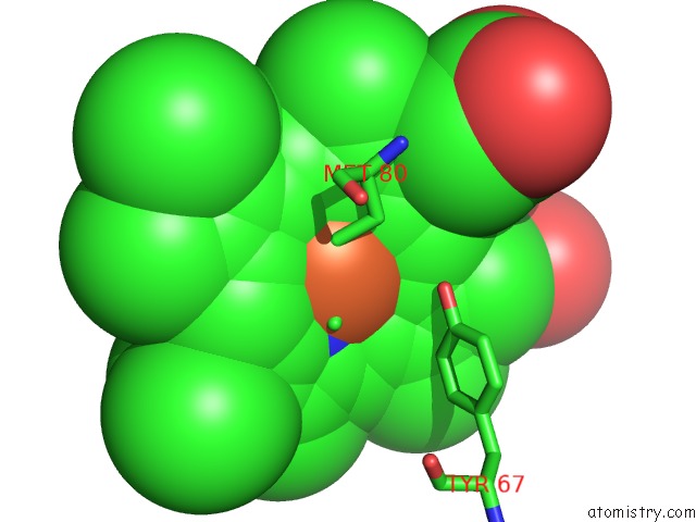
Mono view
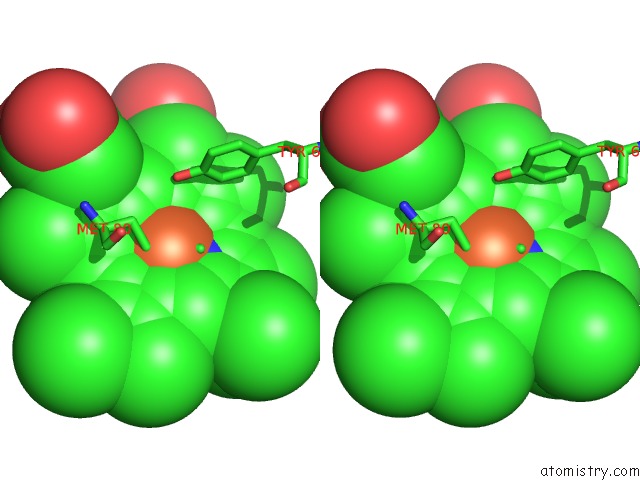
Stereo pair view
Mono view
Stereo pair view
A full contact list of Iron with other atoms in the Fe binding
site number 1 of Cytochrome C Isozyme 1, Reduced, Mutant with Asn 52 Replaced By Ala and Cys 102 Replaced By Thr within 5.0Å range:
|
Reference:
S.P.Rafferty,
J.G.Guillemette,
A.M.Berghuis,
M.Smith,
G.D.Brayer,
A.G.Mauk.
Mechanistic and Structural Contributions of Critical Surface and Internal Residues to Cytochrome C Electron Transfer Reactivity. Biochemistry V. 35 10784 1996.
ISSN: ISSN 0006-2960
PubMed: 8718869
DOI: 10.1021/BI960430V
Page generated: Sat Aug 3 08:12:39 2024
ISSN: ISSN 0006-2960
PubMed: 8718869
DOI: 10.1021/BI960430V
Last articles
Zn in 9J0NZn in 9J0O
Zn in 9J0P
Zn in 9FJX
Zn in 9EKB
Zn in 9C0F
Zn in 9CAH
Zn in 9CH0
Zn in 9CH3
Zn in 9CH1