Iron »
PDB 1iro-1j3y »
1it2 »
Iron in PDB 1it2: Hagfish Deoxy Hemoglobin
Protein crystallography data
The structure of Hagfish Deoxy Hemoglobin, PDB code: 1it2
was solved by
M.Mito,
K.T.Chong,
S.-Y.Park,
J.R.Tame,
with X-Ray Crystallography technique. A brief refinement statistics is given in the table below:
| Resolution Low / High (Å) | 20.00 / 1.60 |
| Space group | C 1 2 1 |
| Cell size a, b, c (Å), α, β, γ (°) | 104.024, 62.269, 66.536, 90.00, 107.40, 90.00 |
| R / Rfree (%) | 21.1 / 24.1 |
Iron Binding Sites:
The binding sites of Iron atom in the Hagfish Deoxy Hemoglobin
(pdb code 1it2). This binding sites where shown within
5.0 Angstroms radius around Iron atom.
In total 2 binding sites of Iron where determined in the Hagfish Deoxy Hemoglobin, PDB code: 1it2:
Jump to Iron binding site number: 1; 2;
In total 2 binding sites of Iron where determined in the Hagfish Deoxy Hemoglobin, PDB code: 1it2:
Jump to Iron binding site number: 1; 2;
Iron binding site 1 out of 2 in 1it2
Go back to
Iron binding site 1 out
of 2 in the Hagfish Deoxy Hemoglobin
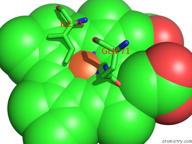
Mono view
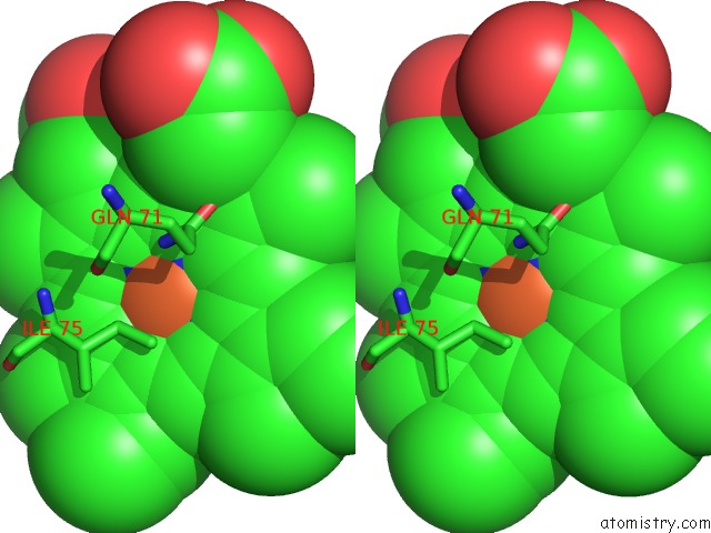
Stereo pair view
Mono view
Stereo pair view
A full contact list of Iron with other atoms in the Fe binding
site number 1 of Hagfish Deoxy Hemoglobin within 5.0Å range:
|
Iron binding site 2 out of 2 in 1it2
Go back to
Iron binding site 2 out
of 2 in the Hagfish Deoxy Hemoglobin
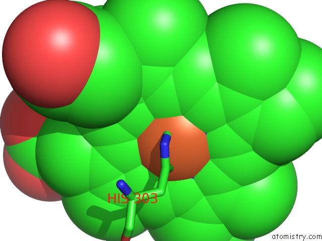
Mono view
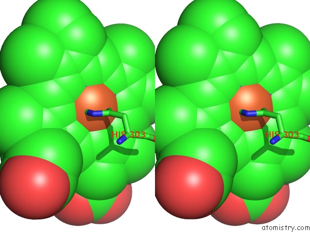
Stereo pair view
Mono view
Stereo pair view
A full contact list of Iron with other atoms in the Fe binding
site number 2 of Hagfish Deoxy Hemoglobin within 5.0Å range:
|
Reference:
M.Mito,
K.T.Chong,
G.Miyazaki,
S.Adachi,
S.-Y.Park,
J.R.Tame,
H.Morimoto.
Crystal Structures of Deoxy- and Carbonmonoxyhemoglobin F1 From the Hagfish Eptatretus Burgeri J.Biol.Chem. V. 277 21898 2002.
ISSN: ISSN 0021-9258
PubMed: 11923284
DOI: 10.1074/JBC.M111492200
Page generated: Wed Jul 16 16:20:06 2025
ISSN: ISSN 0021-9258
PubMed: 11923284
DOI: 10.1074/JBC.M111492200
Last articles
Fe in 2YXOFe in 2YRS
Fe in 2YXC
Fe in 2YNM
Fe in 2YVJ
Fe in 2YP1
Fe in 2YU2
Fe in 2YU1
Fe in 2YQB
Fe in 2YOO