Iron »
PDB 1iro-1j3y »
1iw1 »
Iron in PDB 1iw1: Crystal Structure of A Heme Oxygenase (Hmuo) From Corynebacterium Diphtheriae Complexed with Heme in the Ferrous State
Enzymatic activity of Crystal Structure of A Heme Oxygenase (Hmuo) From Corynebacterium Diphtheriae Complexed with Heme in the Ferrous State
All present enzymatic activity of Crystal Structure of A Heme Oxygenase (Hmuo) From Corynebacterium Diphtheriae Complexed with Heme in the Ferrous State:
1.14.99.3;
1.14.99.3;
Protein crystallography data
The structure of Crystal Structure of A Heme Oxygenase (Hmuo) From Corynebacterium Diphtheriae Complexed with Heme in the Ferrous State, PDB code: 1iw1
was solved by
S.Hirotsu,
M.Unno,
G.C.Chu,
D.S.Lee,
S.Y.Park,
Y.Shiro,
M.Ikeda-Saito,
Riken Structural Genomics/Proteomics Initiative (Rsgi),
with X-Ray Crystallography technique. A brief refinement statistics is given in the table below:
| Resolution Low / High (Å) | 12.00 / 1.50 |
| Space group | P 1 21 1 |
| Cell size a, b, c (Å), α, β, γ (°) | 53.865, 62.837, 107.208, 90.00, 101.02, 90.00 |
| R / Rfree (%) | 17.7 / 20.2 |
Iron Binding Sites:
The binding sites of Iron atom in the Crystal Structure of A Heme Oxygenase (Hmuo) From Corynebacterium Diphtheriae Complexed with Heme in the Ferrous State
(pdb code 1iw1). This binding sites where shown within
5.0 Angstroms radius around Iron atom.
In total 3 binding sites of Iron where determined in the Crystal Structure of A Heme Oxygenase (Hmuo) From Corynebacterium Diphtheriae Complexed with Heme in the Ferrous State, PDB code: 1iw1:
Jump to Iron binding site number: 1; 2; 3;
In total 3 binding sites of Iron where determined in the Crystal Structure of A Heme Oxygenase (Hmuo) From Corynebacterium Diphtheriae Complexed with Heme in the Ferrous State, PDB code: 1iw1:
Jump to Iron binding site number: 1; 2; 3;
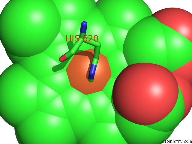
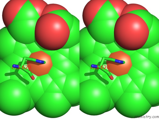
Iron binding site 1 out of 3 in 1iw1
Go back to
Iron binding site 1 out
of 3 in the Crystal Structure of A Heme Oxygenase (Hmuo) From Corynebacterium Diphtheriae Complexed with Heme in the Ferrous State
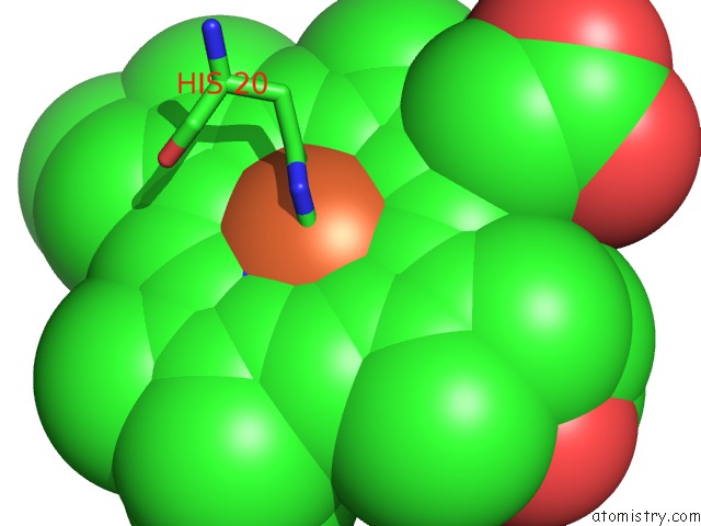
Mono view
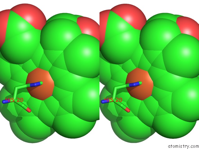
Stereo pair view
Mono view
Stereo pair view
A full contact list of Iron with other atoms in the Fe binding
site number 1 of Crystal Structure of A Heme Oxygenase (Hmuo) From Corynebacterium Diphtheriae Complexed with Heme in the Ferrous State within 5.0Å range:
|
Iron binding site 2 out of 3 in 1iw1
Go back to
Iron binding site 2 out
of 3 in the Crystal Structure of A Heme Oxygenase (Hmuo) From Corynebacterium Diphtheriae Complexed with Heme in the Ferrous State
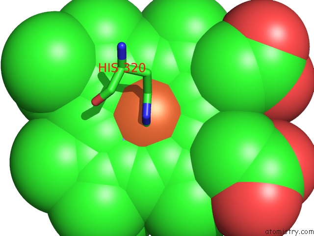
Mono view
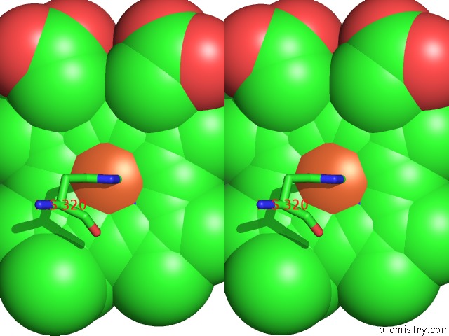
Stereo pair view
Mono view
Stereo pair view
A full contact list of Iron with other atoms in the Fe binding
site number 2 of Crystal Structure of A Heme Oxygenase (Hmuo) From Corynebacterium Diphtheriae Complexed with Heme in the Ferrous State within 5.0Å range:
|
Iron binding site 3 out of 3 in 1iw1
Go back to
Iron binding site 3 out
of 3 in the Crystal Structure of A Heme Oxygenase (Hmuo) From Corynebacterium Diphtheriae Complexed with Heme in the Ferrous State
Mono view
Stereo pair view
Mono view
Stereo pair view
A full contact list of Iron with other atoms in the Fe binding
site number 3 of Crystal Structure of A Heme Oxygenase (Hmuo) From Corynebacterium Diphtheriae Complexed with Heme in the Ferrous State within 5.0Å range:
|
Reference:
S.Hirotsu,
G.C.Chu,
M.Unno,
D.S.Lee,
T.Yoshida,
S.Y.Park,
Y.Shiro,
M.Ikeda-Saito.
The Crystal Structures of the Ferric and Ferrous Forms of the Heme Complex of Hmuo, A Heme Oxygenase of Corynebacterium Diphtheriae. J.Biol.Chem. V. 279 11937 2004.
ISSN: ISSN 0021-9258
PubMed: 14645223
DOI: 10.1074/JBC.M311631200
Page generated: Sat Aug 3 08:15:47 2024
ISSN: ISSN 0021-9258
PubMed: 14645223
DOI: 10.1074/JBC.M311631200
Last articles
Zn in 9J0NZn in 9J0O
Zn in 9J0P
Zn in 9FJX
Zn in 9EKB
Zn in 9C0F
Zn in 9CAH
Zn in 9CH0
Zn in 9CH3
Zn in 9CH1