Iron »
PDB 1iro-1j3y »
1j2c »
Iron in PDB 1j2c: Crystal Structure of Rat Heme Oxygenase-1 in Complex with Biliverdin Ixalpha-Iron Cluster
Enzymatic activity of Crystal Structure of Rat Heme Oxygenase-1 in Complex with Biliverdin Ixalpha-Iron Cluster
All present enzymatic activity of Crystal Structure of Rat Heme Oxygenase-1 in Complex with Biliverdin Ixalpha-Iron Cluster:
1.14.99.3;
1.14.99.3;
Protein crystallography data
The structure of Crystal Structure of Rat Heme Oxygenase-1 in Complex with Biliverdin Ixalpha-Iron Cluster, PDB code: 1j2c
was solved by
M.Sugishima,
H.Sakamoto,
M.Noguchi,
K.Fukuyama,
with X-Ray Crystallography technique. A brief refinement statistics is given in the table below:
| Resolution Low / High (Å) | 60.80 / 2.40 |
| Space group | P 32 2 1 |
| Cell size a, b, c (Å), α, β, γ (°) | 65.400, 65.400, 121.300, 90.00, 90.00, 120.00 |
| R / Rfree (%) | 19.4 / 23.9 |
Iron Binding Sites:
The binding sites of Iron atom in the Crystal Structure of Rat Heme Oxygenase-1 in Complex with Biliverdin Ixalpha-Iron Cluster
(pdb code 1j2c). This binding sites where shown within
5.0 Angstroms radius around Iron atom.
In total only one binding site of Iron was determined in the Crystal Structure of Rat Heme Oxygenase-1 in Complex with Biliverdin Ixalpha-Iron Cluster, PDB code: 1j2c:
In total only one binding site of Iron was determined in the Crystal Structure of Rat Heme Oxygenase-1 in Complex with Biliverdin Ixalpha-Iron Cluster, PDB code: 1j2c:
Iron binding site 1 out of 1 in 1j2c
Go back to
Iron binding site 1 out
of 1 in the Crystal Structure of Rat Heme Oxygenase-1 in Complex with Biliverdin Ixalpha-Iron Cluster
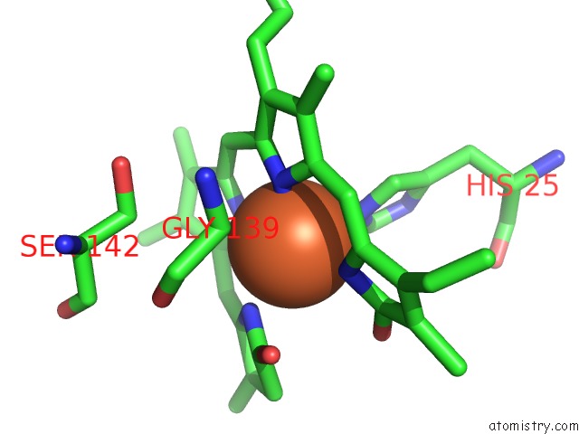
Mono view
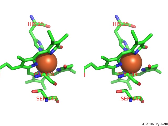
Stereo pair view
Mono view
Stereo pair view
A full contact list of Iron with other atoms in the Fe binding
site number 1 of Crystal Structure of Rat Heme Oxygenase-1 in Complex with Biliverdin Ixalpha-Iron Cluster within 5.0Å range:
|
Reference:
M.Sugishima,
H.Sakamoto,
Y.Higashimoto,
M.Noguchi,
K.Fukuyama.
Crystal Structure of Rat Heme Oxygenase-1 in Complex with Biliverdin-Iron Chelate: Conformational Change of the Distal Helix During the Heme Cleavage Reaction. J.Biol.Chem. V. 278 32352 2003.
ISSN: ISSN 0021-9258
PubMed: 12794075
DOI: 10.1074/JBC.M303682200
Page generated: Sat Aug 3 08:19:39 2024
ISSN: ISSN 0021-9258
PubMed: 12794075
DOI: 10.1074/JBC.M303682200
Last articles
Zn in 9J0NZn in 9J0O
Zn in 9J0P
Zn in 9FJX
Zn in 9EKB
Zn in 9C0F
Zn in 9CAH
Zn in 9CH0
Zn in 9CH3
Zn in 9CH1