Iron »
PDB 1iro-1j3y »
1j30 »
Iron in PDB 1j30: The Crystal Structure of Sulerythrin, A Rubrerythrin-Like Protein From A Strictly Aerobic and Thermoacidiphilic Archaeon
Protein crystallography data
The structure of The Crystal Structure of Sulerythrin, A Rubrerythrin-Like Protein From A Strictly Aerobic and Thermoacidiphilic Archaeon, PDB code: 1j30
was solved by
S.Fushinobu,
H.Shoun,
T.Wakagi,
with X-Ray Crystallography technique. A brief refinement statistics is given in the table below:
| Resolution Low / High (Å) | 24.29 / 1.70 |
| Space group | P 63 |
| Cell size a, b, c (Å), α, β, γ (°) | 72.428, 72.428, 98.208, 90.00, 90.00, 120.00 |
| R / Rfree (%) | 19.2 / 22.3 |
Other elements in 1j30:
The structure of The Crystal Structure of Sulerythrin, A Rubrerythrin-Like Protein From A Strictly Aerobic and Thermoacidiphilic Archaeon also contains other interesting chemical elements:
| Zinc | (Zn) | 2 atoms |
Iron Binding Sites:
The binding sites of Iron atom in the The Crystal Structure of Sulerythrin, A Rubrerythrin-Like Protein From A Strictly Aerobic and Thermoacidiphilic Archaeon
(pdb code 1j30). This binding sites where shown within
5.0 Angstroms radius around Iron atom.
In total 2 binding sites of Iron where determined in the The Crystal Structure of Sulerythrin, A Rubrerythrin-Like Protein From A Strictly Aerobic and Thermoacidiphilic Archaeon, PDB code: 1j30:
Jump to Iron binding site number: 1; 2;
In total 2 binding sites of Iron where determined in the The Crystal Structure of Sulerythrin, A Rubrerythrin-Like Protein From A Strictly Aerobic and Thermoacidiphilic Archaeon, PDB code: 1j30:
Jump to Iron binding site number: 1; 2;
Iron binding site 1 out of 2 in 1j30
Go back to
Iron binding site 1 out
of 2 in the The Crystal Structure of Sulerythrin, A Rubrerythrin-Like Protein From A Strictly Aerobic and Thermoacidiphilic Archaeon
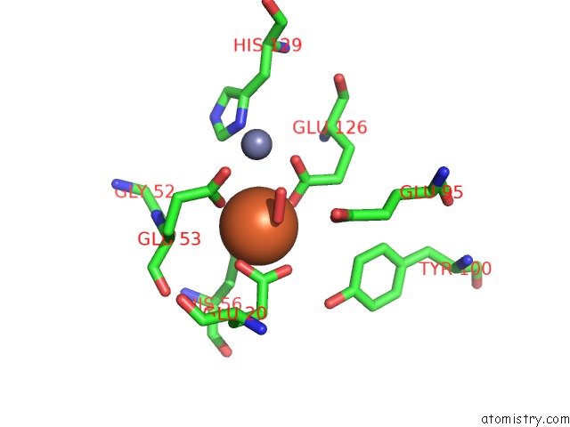
Mono view
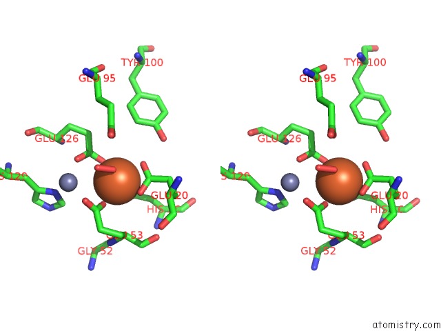
Stereo pair view
Mono view
Stereo pair view
A full contact list of Iron with other atoms in the Fe binding
site number 1 of The Crystal Structure of Sulerythrin, A Rubrerythrin-Like Protein From A Strictly Aerobic and Thermoacidiphilic Archaeon within 5.0Å range:
|
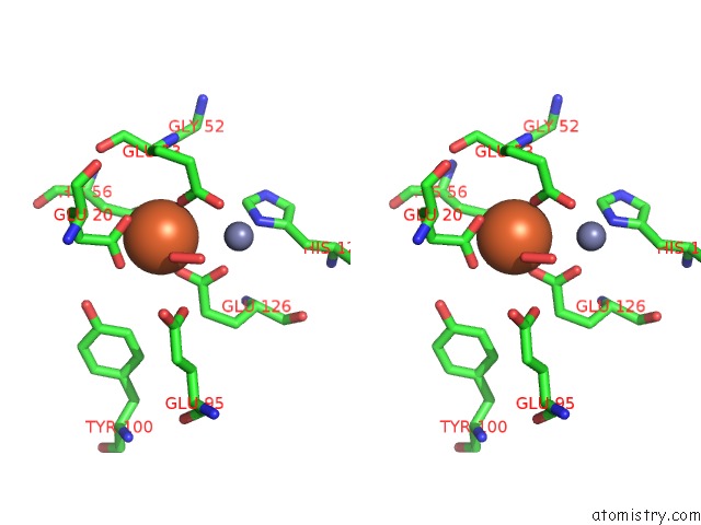
Iron binding site 2 out of 2 in 1j30
Go back to
Iron binding site 2 out
of 2 in the The Crystal Structure of Sulerythrin, A Rubrerythrin-Like Protein From A Strictly Aerobic and Thermoacidiphilic Archaeon
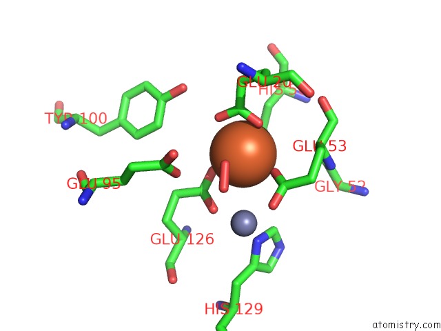
Mono view
Stereo pair view
Mono view
Stereo pair view
A full contact list of Iron with other atoms in the Fe binding
site number 2 of The Crystal Structure of Sulerythrin, A Rubrerythrin-Like Protein From A Strictly Aerobic and Thermoacidiphilic Archaeon within 5.0Å range:
|
Reference:
S.Fushinobu,
H.Shoun,
T.Wakagi.
The Crystal Structure of Sulerythrin, A Rubrerythrin-Like Protein From A Strictly Aerobic Archaeon, Sulfolobus Tokodaii Strain 7, Shows Unexpected Domain Swapping Biochemistry V. 42 11707 2003.
ISSN: ISSN 0006-2960
PubMed: 14529281
DOI: 10.1021/BI034220B
Page generated: Sat Aug 3 08:19:43 2024
ISSN: ISSN 0006-2960
PubMed: 14529281
DOI: 10.1021/BI034220B
Last articles
Zn in 9J0NZn in 9J0O
Zn in 9J0P
Zn in 9FJX
Zn in 9EKB
Zn in 9C0F
Zn in 9CAH
Zn in 9CH0
Zn in 9CH3
Zn in 9CH1