Iron »
PDB 1j3z-1jju »
1j40 »
Iron in PDB 1j40: Direct Observation of Photolysis-Induced Tertiary Structural Changes in Human Haemoglobin; Crystal Structure of Alpha(Ni)-Beta(Fe-Co) Hemoglobin (Laser Unphotolysed)
Protein crystallography data
The structure of Direct Observation of Photolysis-Induced Tertiary Structural Changes in Human Haemoglobin; Crystal Structure of Alpha(Ni)-Beta(Fe-Co) Hemoglobin (Laser Unphotolysed), PDB code: 1j40
was solved by
S.Adachi,
S.-Y.Park,
J.R.H.Tame,
Y.Shiro,
N.Shibayama,
Rikenstructural Genomics/Proteomics Initiative (Rsgi),
with X-Ray Crystallography technique. A brief refinement statistics is given in the table below:
| Resolution Low / High (Å) | 20.00 / 1.45 |
| Space group | P 1 21 1 |
| Cell size a, b, c (Å), α, β, γ (°) | 64.708, 94.860, 99.730, 90.00, 101.97, 90.00 |
| R / Rfree (%) | 16.5 / 19.7 |
Other elements in 1j40:
The structure of Direct Observation of Photolysis-Induced Tertiary Structural Changes in Human Haemoglobin; Crystal Structure of Alpha(Ni)-Beta(Fe-Co) Hemoglobin (Laser Unphotolysed) also contains other interesting chemical elements:
| Nickel | (Ni) | 4 atoms |
Iron Binding Sites:
The binding sites of Iron atom in the Direct Observation of Photolysis-Induced Tertiary Structural Changes in Human Haemoglobin; Crystal Structure of Alpha(Ni)-Beta(Fe-Co) Hemoglobin (Laser Unphotolysed)
(pdb code 1j40). This binding sites where shown within
5.0 Angstroms radius around Iron atom.
In total 4 binding sites of Iron where determined in the Direct Observation of Photolysis-Induced Tertiary Structural Changes in Human Haemoglobin; Crystal Structure of Alpha(Ni)-Beta(Fe-Co) Hemoglobin (Laser Unphotolysed), PDB code: 1j40:
Jump to Iron binding site number: 1; 2; 3; 4;
In total 4 binding sites of Iron where determined in the Direct Observation of Photolysis-Induced Tertiary Structural Changes in Human Haemoglobin; Crystal Structure of Alpha(Ni)-Beta(Fe-Co) Hemoglobin (Laser Unphotolysed), PDB code: 1j40:
Jump to Iron binding site number: 1; 2; 3; 4;
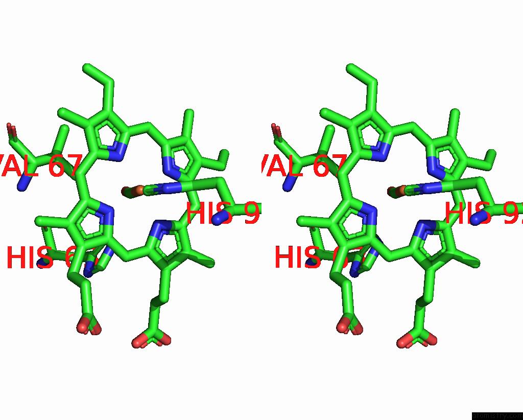
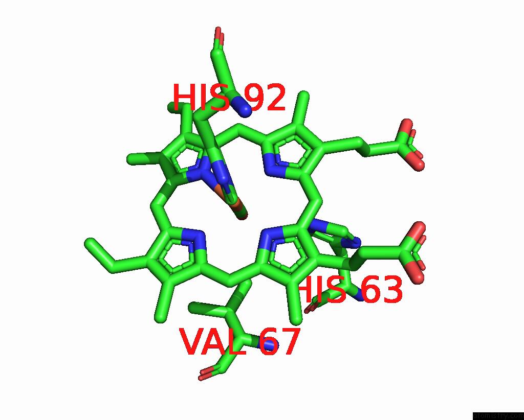
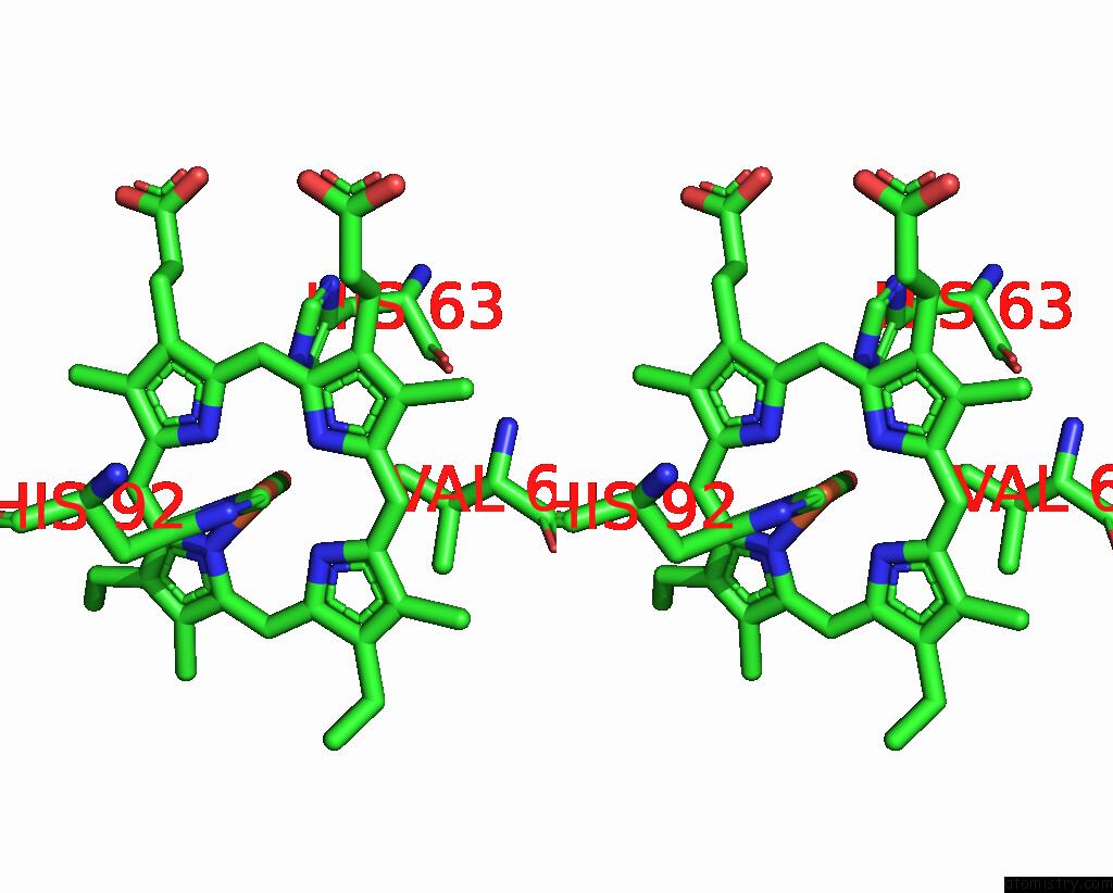
Iron binding site 1 out of 4 in 1j40
Go back to
Iron binding site 1 out
of 4 in the Direct Observation of Photolysis-Induced Tertiary Structural Changes in Human Haemoglobin; Crystal Structure of Alpha(Ni)-Beta(Fe-Co) Hemoglobin (Laser Unphotolysed)
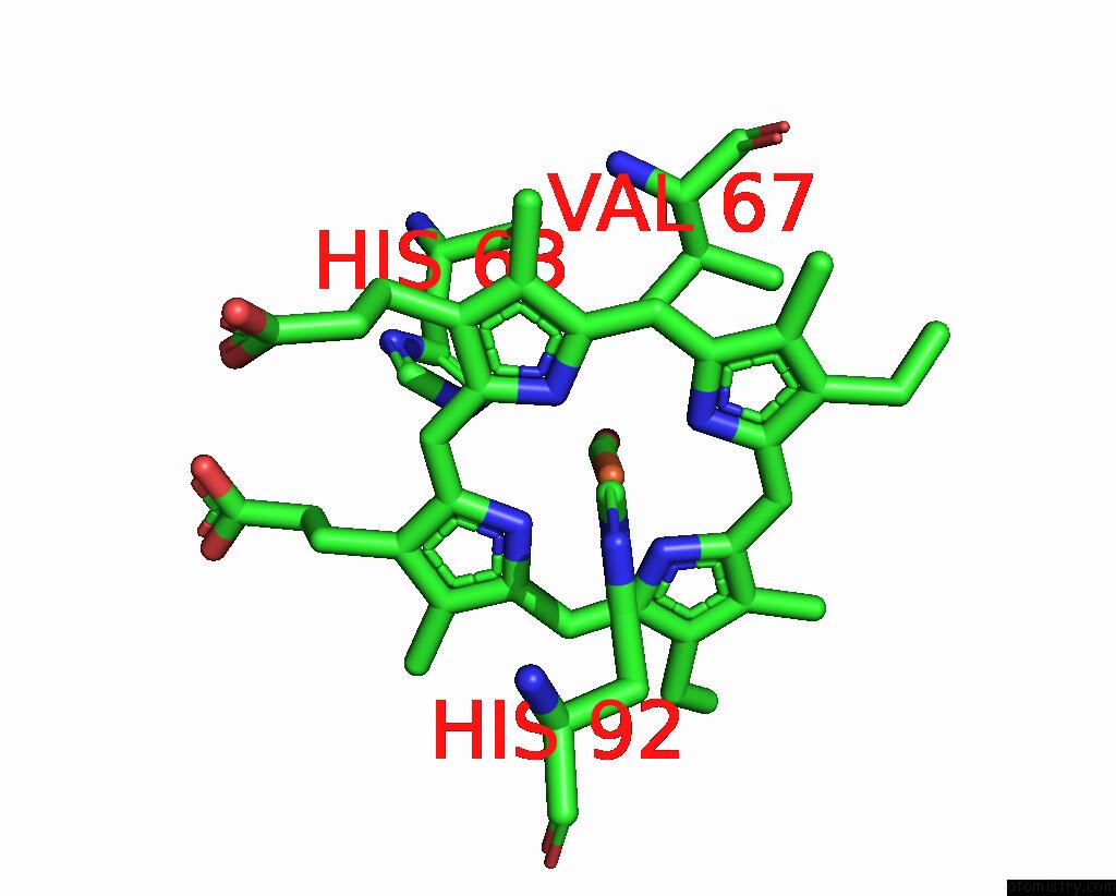
Mono view
Stereo pair view
Mono view
Stereo pair view
A full contact list of Iron with other atoms in the Fe binding
site number 1 of Direct Observation of Photolysis-Induced Tertiary Structural Changes in Human Haemoglobin; Crystal Structure of Alpha(Ni)-Beta(Fe-Co) Hemoglobin (Laser Unphotolysed) within 5.0Å range:
|
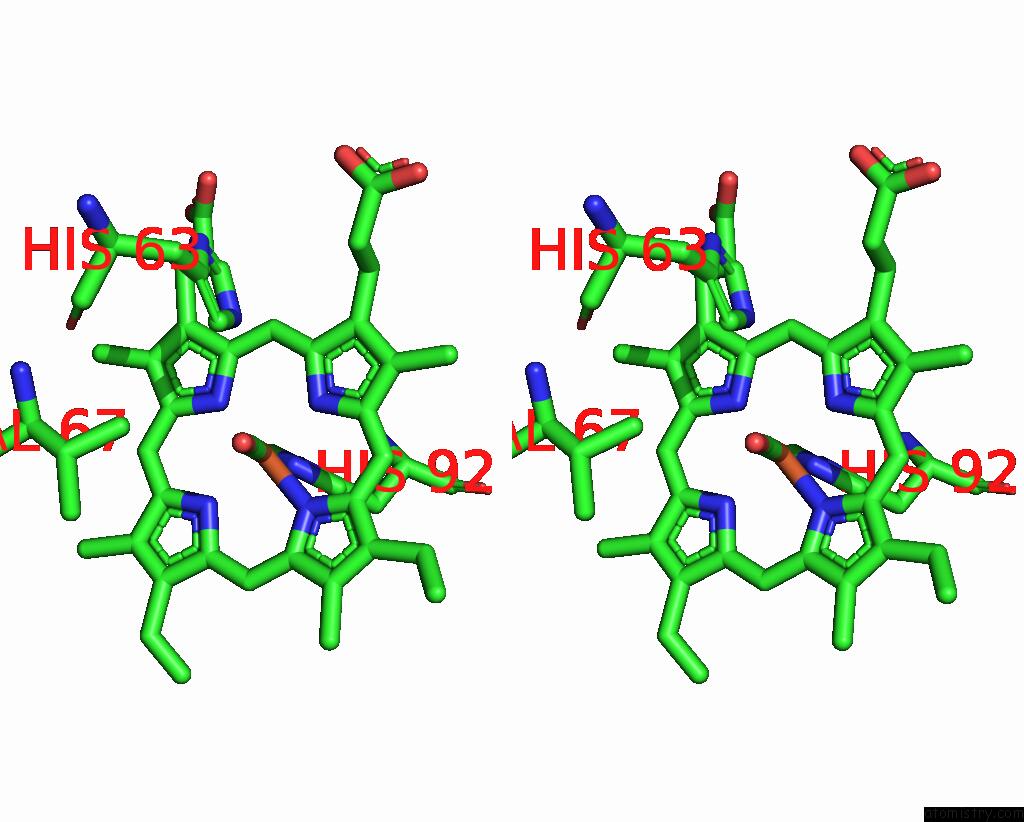
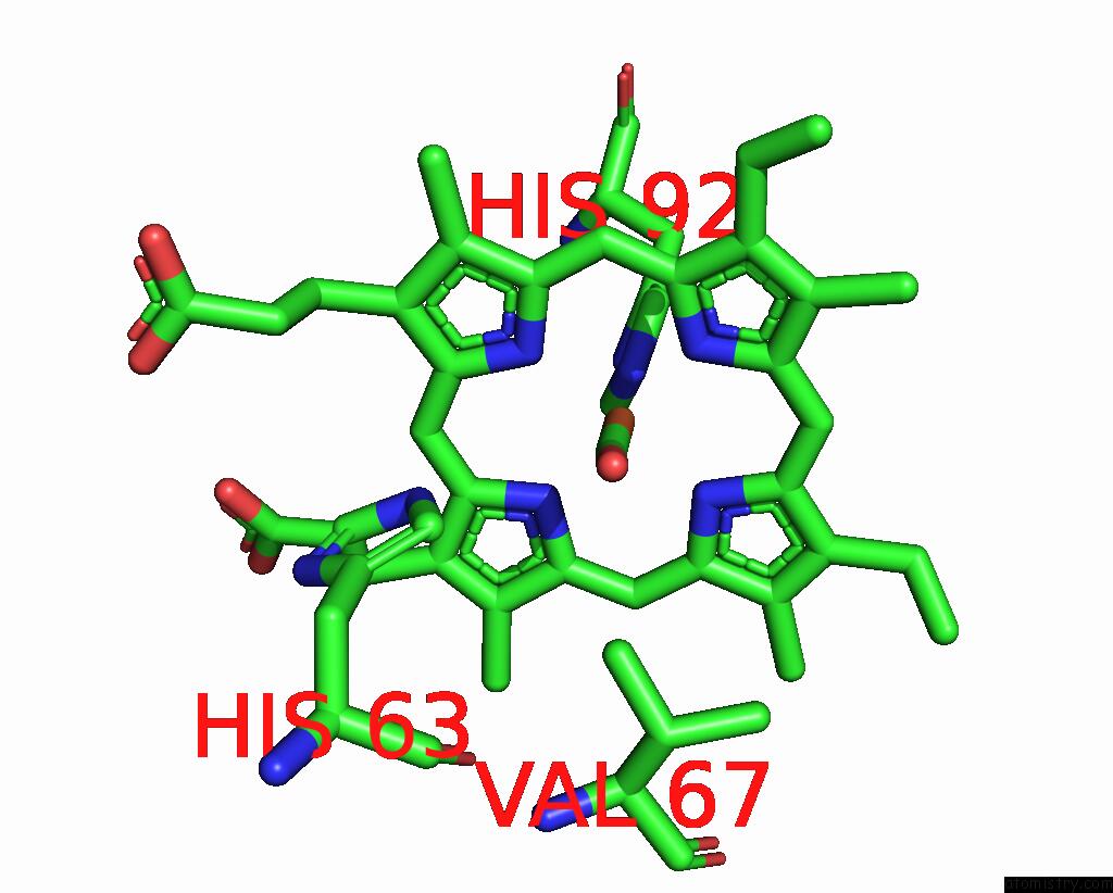
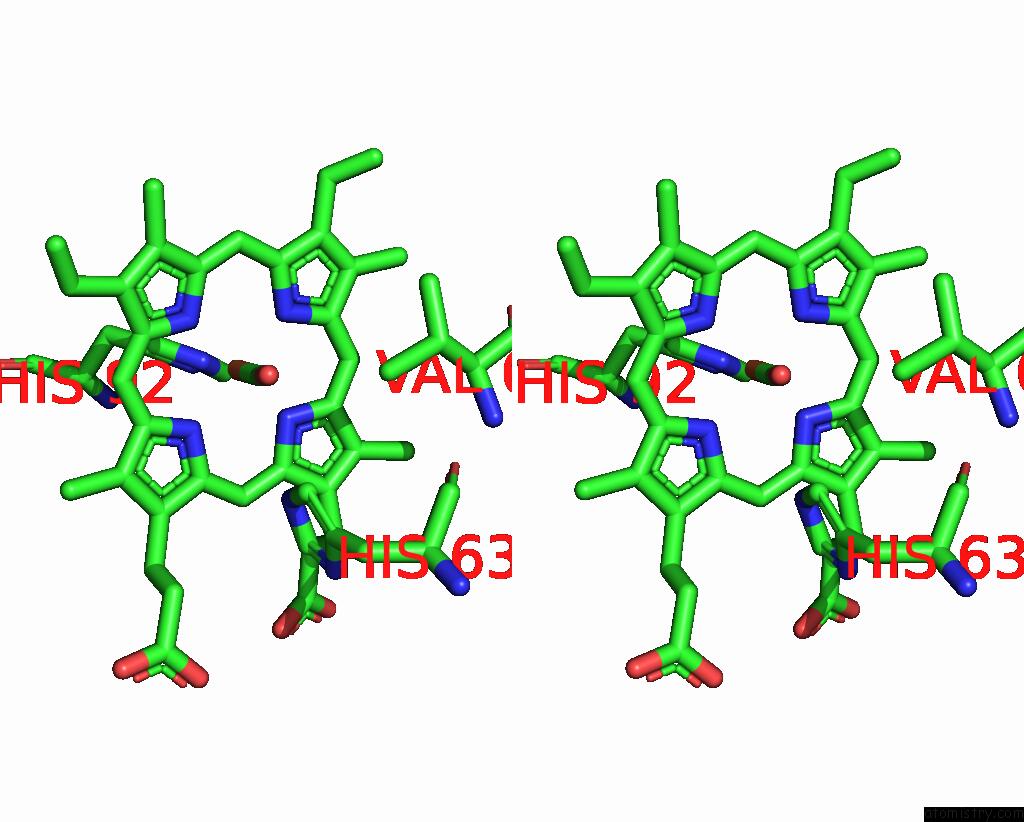
Iron binding site 2 out of 4 in 1j40
Go back to
Iron binding site 2 out
of 4 in the Direct Observation of Photolysis-Induced Tertiary Structural Changes in Human Haemoglobin; Crystal Structure of Alpha(Ni)-Beta(Fe-Co) Hemoglobin (Laser Unphotolysed)
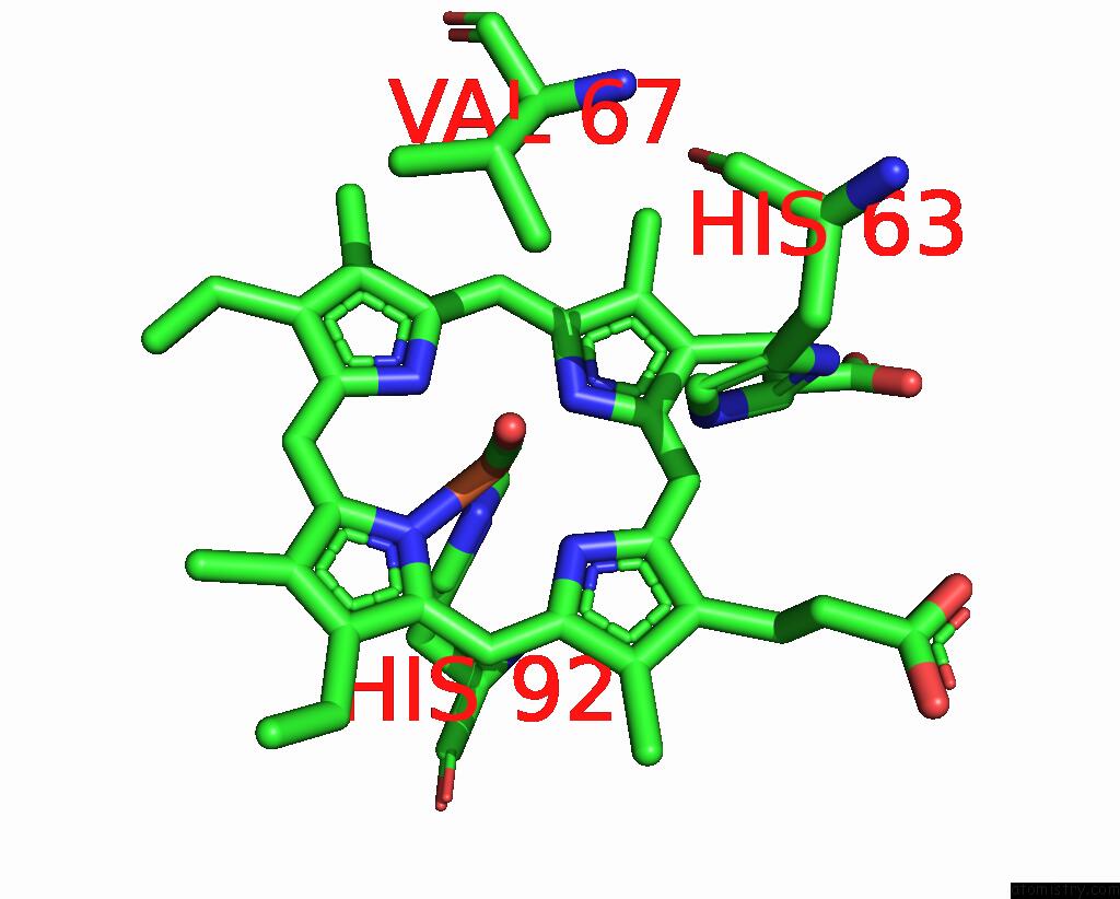
Mono view
Stereo pair view
Mono view
Stereo pair view
A full contact list of Iron with other atoms in the Fe binding
site number 2 of Direct Observation of Photolysis-Induced Tertiary Structural Changes in Human Haemoglobin; Crystal Structure of Alpha(Ni)-Beta(Fe-Co) Hemoglobin (Laser Unphotolysed) within 5.0Å range:
|
Iron binding site 3 out of 4 in 1j40
Go back to
Iron binding site 3 out
of 4 in the Direct Observation of Photolysis-Induced Tertiary Structural Changes in Human Haemoglobin; Crystal Structure of Alpha(Ni)-Beta(Fe-Co) Hemoglobin (Laser Unphotolysed)
Mono view
Stereo pair view
Mono view
Stereo pair view
A full contact list of Iron with other atoms in the Fe binding
site number 3 of Direct Observation of Photolysis-Induced Tertiary Structural Changes in Human Haemoglobin; Crystal Structure of Alpha(Ni)-Beta(Fe-Co) Hemoglobin (Laser Unphotolysed) within 5.0Å range:
|
Iron binding site 4 out of 4 in 1j40
Go back to
Iron binding site 4 out
of 4 in the Direct Observation of Photolysis-Induced Tertiary Structural Changes in Human Haemoglobin; Crystal Structure of Alpha(Ni)-Beta(Fe-Co) Hemoglobin (Laser Unphotolysed)
Mono view
Stereo pair view
Mono view
Stereo pair view
A full contact list of Iron with other atoms in the Fe binding
site number 4 of Direct Observation of Photolysis-Induced Tertiary Structural Changes in Human Haemoglobin; Crystal Structure of Alpha(Ni)-Beta(Fe-Co) Hemoglobin (Laser Unphotolysed) within 5.0Å range:
|
Reference:
S.Adachi,
S.-Y.Park,
J.R.H.Tame,
Y.Shiro,
N.Shibayama.
Direct Observation of Photolysis-Induced Tertiary Structural Changes in Hemoglobin Proc.Natl.Acad.Sci.Usa V. 100 7039 2003.
ISSN: ISSN 0027-8424
PubMed: 12773618
DOI: 10.1073/PNAS.1230629100
Page generated: Sat Aug 3 08:23:05 2024
ISSN: ISSN 0027-8424
PubMed: 12773618
DOI: 10.1073/PNAS.1230629100
Last articles
Zn in 9MJ5Zn in 9HNW
Zn in 9G0L
Zn in 9FNE
Zn in 9DZN
Zn in 9E0I
Zn in 9D32
Zn in 9DAK
Zn in 8ZXC
Zn in 8ZUF