Iron »
PDB 1j3z-1jju »
1j7w »
Iron in PDB 1j7w: Crystal Structure of Deoxy Hbbetayq, A Site Directed Mutant of Hba
Protein crystallography data
The structure of Crystal Structure of Deoxy Hbbetayq, A Site Directed Mutant of Hba, PDB code: 1j7w
was solved by
A.E.Miele,
F.Draghi,
A.Arcovito,
A.Bellelli,
M.Brunori,
C.Travaglini-Allocatelli,
B.Vallone,
with X-Ray Crystallography technique. A brief refinement statistics is given in the table below:
| Resolution Low / High (Å) | 29.00 / 2.00 |
| Space group | P 1 21 1 |
| Cell size a, b, c (Å), α, β, γ (°) | 63.360, 84.320, 54.000, 90.00, 99.43, 90.00 |
| R / Rfree (%) | 18.1 / 22.3 |
Iron Binding Sites:
The binding sites of Iron atom in the Crystal Structure of Deoxy Hbbetayq, A Site Directed Mutant of Hba
(pdb code 1j7w). This binding sites where shown within
5.0 Angstroms radius around Iron atom.
In total 4 binding sites of Iron where determined in the Crystal Structure of Deoxy Hbbetayq, A Site Directed Mutant of Hba, PDB code: 1j7w:
Jump to Iron binding site number: 1; 2; 3; 4;
In total 4 binding sites of Iron where determined in the Crystal Structure of Deoxy Hbbetayq, A Site Directed Mutant of Hba, PDB code: 1j7w:
Jump to Iron binding site number: 1; 2; 3; 4;
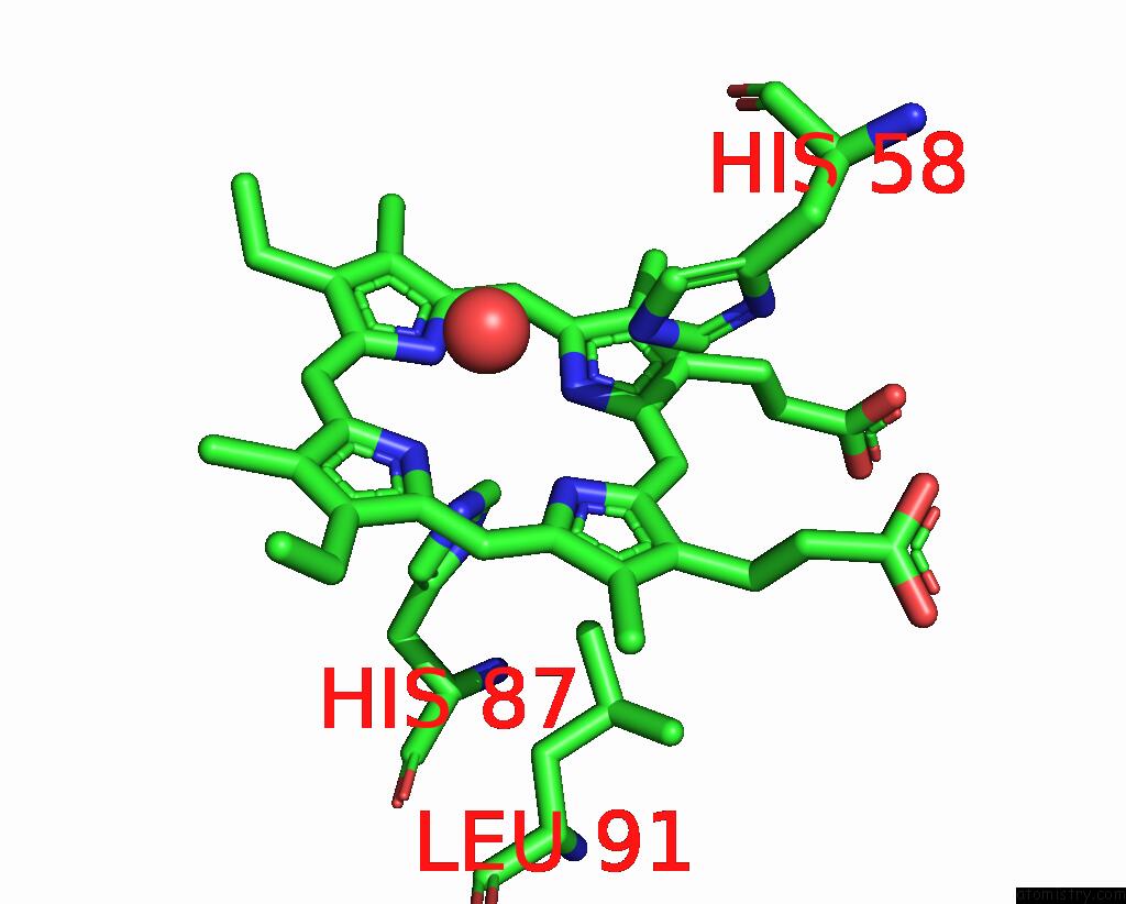
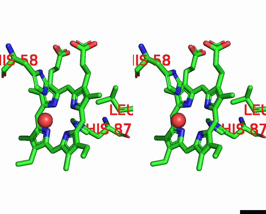
Iron binding site 1 out of 4 in 1j7w
Go back to
Iron binding site 1 out
of 4 in the Crystal Structure of Deoxy Hbbetayq, A Site Directed Mutant of Hba
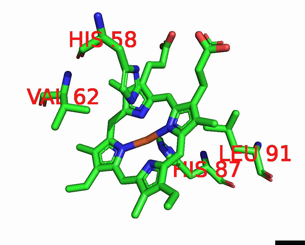
Mono view
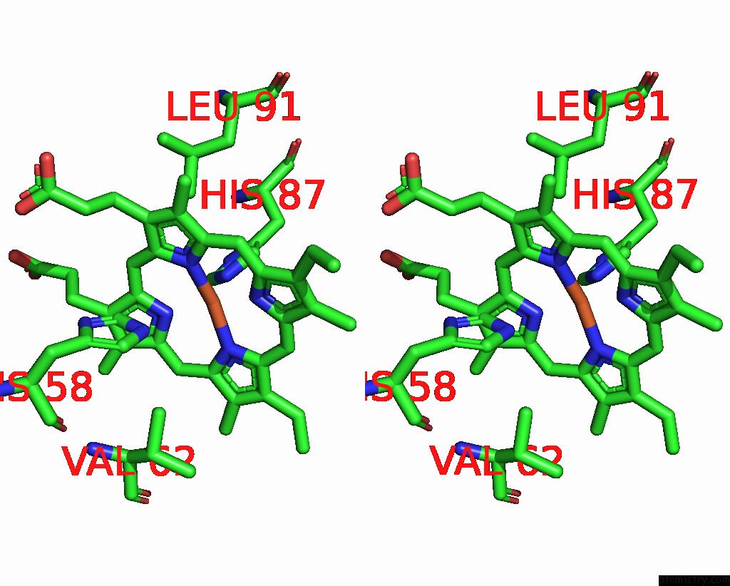
Stereo pair view
Mono view
Stereo pair view
A full contact list of Iron with other atoms in the Fe binding
site number 1 of Crystal Structure of Deoxy Hbbetayq, A Site Directed Mutant of Hba within 5.0Å range:
|
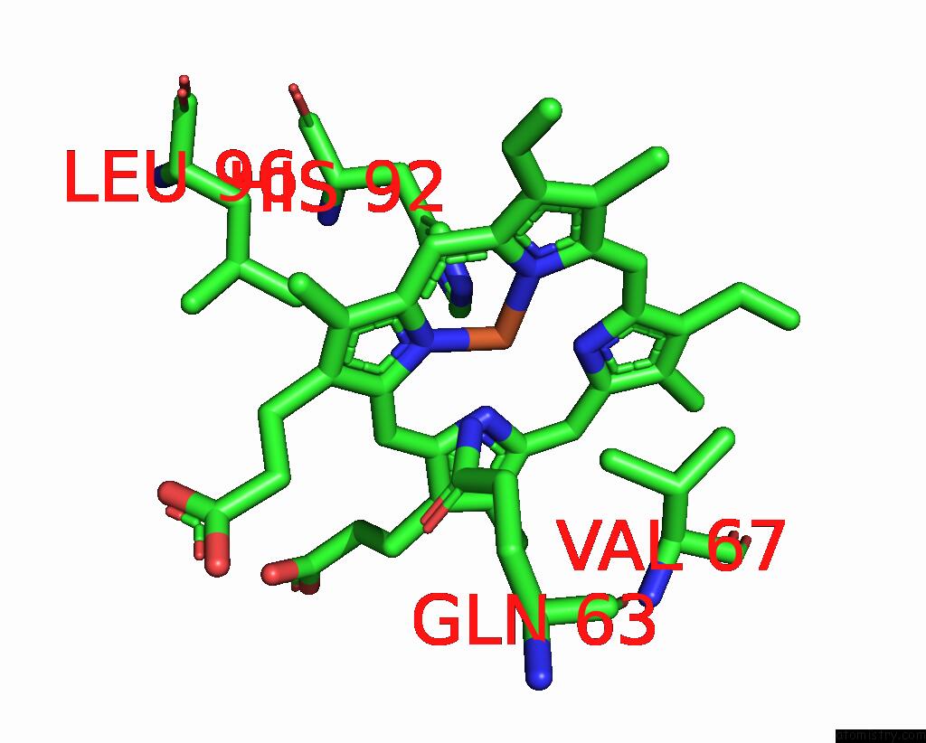
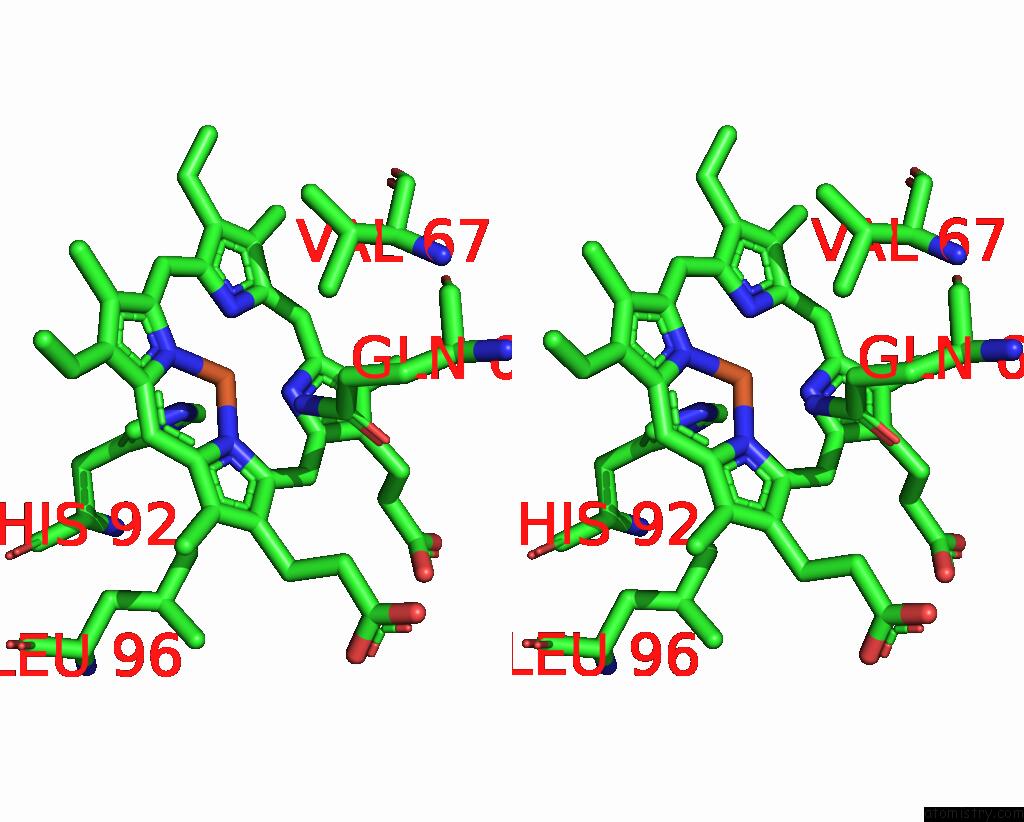
Iron binding site 2 out of 4 in 1j7w
Go back to
Iron binding site 2 out
of 4 in the Crystal Structure of Deoxy Hbbetayq, A Site Directed Mutant of Hba
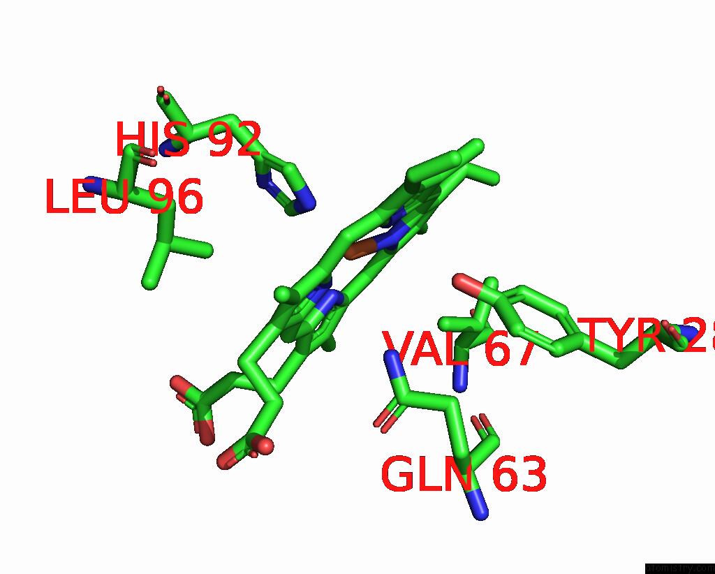
Mono view
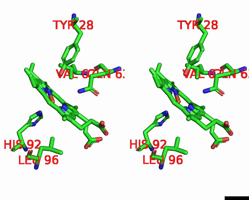
Stereo pair view
Mono view
Stereo pair view
A full contact list of Iron with other atoms in the Fe binding
site number 2 of Crystal Structure of Deoxy Hbbetayq, A Site Directed Mutant of Hba within 5.0Å range:
|
Iron binding site 3 out of 4 in 1j7w
Go back to
Iron binding site 3 out
of 4 in the Crystal Structure of Deoxy Hbbetayq, A Site Directed Mutant of Hba
Mono view
Stereo pair view
Mono view
Stereo pair view
A full contact list of Iron with other atoms in the Fe binding
site number 3 of Crystal Structure of Deoxy Hbbetayq, A Site Directed Mutant of Hba within 5.0Å range:
|
Iron binding site 4 out of 4 in 1j7w
Go back to
Iron binding site 4 out
of 4 in the Crystal Structure of Deoxy Hbbetayq, A Site Directed Mutant of Hba
Mono view
Stereo pair view
Mono view
Stereo pair view
A full contact list of Iron with other atoms in the Fe binding
site number 4 of Crystal Structure of Deoxy Hbbetayq, A Site Directed Mutant of Hba within 5.0Å range:
|
Reference:
A.E.Miele,
F.Draghi,
A.Arcovito,
A.Bellelli,
M.Brunori,
C.Travaglini-Allocatelli,
B.Vallone.
Control of Heme Reactivity By Diffusion: Structural Basis and Functional Characterization in Hemoglobin Mutants. Biochemistry V. 40 14449 2001.
ISSN: ISSN 0006-2960
PubMed: 11724557
DOI: 10.1021/BI011602D
Page generated: Sat Aug 3 08:24:23 2024
ISSN: ISSN 0006-2960
PubMed: 11724557
DOI: 10.1021/BI011602D
Last articles
Cl in 5YQACl in 5YPL
Cl in 5YPK
Cl in 5YPJ
Cl in 5YPI
Cl in 5YPD
Cl in 5YKG
Cl in 5YKF
Cl in 5YKE
Cl in 5YJH