Iron »
PDB 1j3z-1jju »
1jdo »
Iron in PDB 1jdo: Sperm Whale Myoglobin (Ferrous, Nitric Oxide Bound)
Protein crystallography data
The structure of Sperm Whale Myoglobin (Ferrous, Nitric Oxide Bound), PDB code: 1jdo
was solved by
E.A.Brucker,
G.N.Phillips Jr.,
with X-Ray Crystallography technique. A brief refinement statistics is given in the table below:
| Resolution Low / High (Å) | 5.00 / 1.90 |
| Space group | P 6 |
| Cell size a, b, c (Å), α, β, γ (°) | 91.506, 91.506, 45.853, 90.00, 90.00, 120.00 |
| R / Rfree (%) | 15.3 / 20.6 |
Iron Binding Sites:
The binding sites of Iron atom in the Sperm Whale Myoglobin (Ferrous, Nitric Oxide Bound)
(pdb code 1jdo). This binding sites where shown within
5.0 Angstroms radius around Iron atom.
In total only one binding site of Iron was determined in the Sperm Whale Myoglobin (Ferrous, Nitric Oxide Bound), PDB code: 1jdo:
In total only one binding site of Iron was determined in the Sperm Whale Myoglobin (Ferrous, Nitric Oxide Bound), PDB code: 1jdo:
Iron binding site 1 out of 1 in 1jdo
Go back to
Iron binding site 1 out
of 1 in the Sperm Whale Myoglobin (Ferrous, Nitric Oxide Bound)
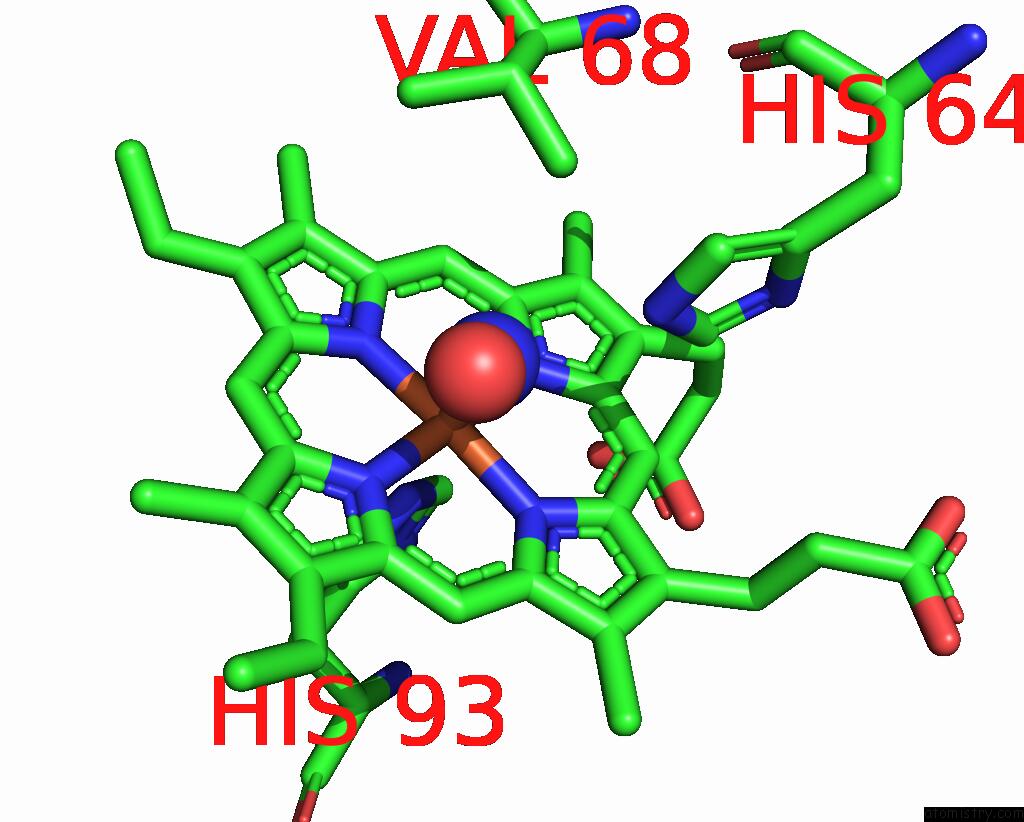
Mono view
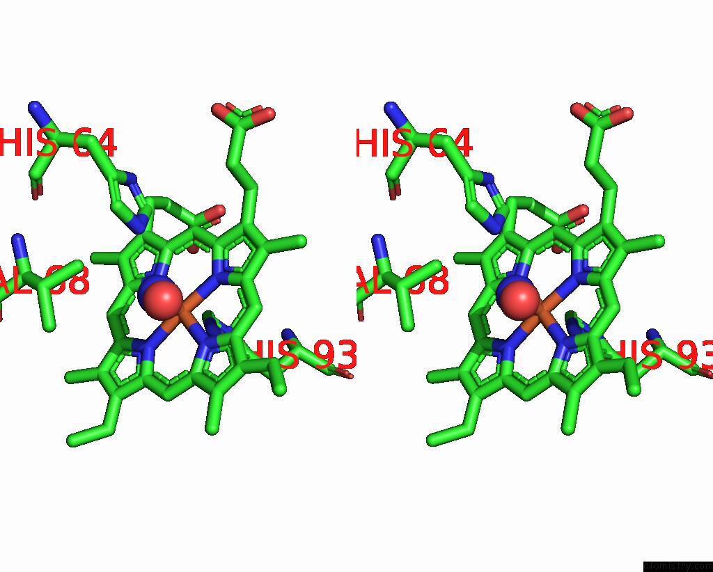
Stereo pair view
Mono view
Stereo pair view
A full contact list of Iron with other atoms in the Fe binding
site number 1 of Sperm Whale Myoglobin (Ferrous, Nitric Oxide Bound) within 5.0Å range:
|
Reference:
E.A.Brucker,
J.S.Olson,
M.Ikeda-Saito,
G.N.Phillips Jr..
Nitric Oxide Myoglobin: Crystal Structure and Analysis of Ligand Geometry. Proteins V. 30 352 1998.
ISSN: ISSN 0887-3585
PubMed: 9533619
DOI: 10.1002/(SICI)1097-0134(19980301)30:4<352::AID-PROT2>3.3.CO;2-G
Page generated: Sat Aug 3 08:27:05 2024
ISSN: ISSN 0887-3585
PubMed: 9533619
DOI: 10.1002/(SICI)1097-0134(19980301)30:4<352::AID-PROT2>3.3.CO;2-G
Last articles
F in 4IVOF in 4IV4
F in 4IVM
F in 4IV2
F in 4IUI
F in 4IN4
F in 4IU7
F in 4ITI
F in 4IUE
F in 4IRU