Iron »
PDB 1j3z-1jju »
1jfb »
Iron in PDB 1jfb: X-Ray Structure of Nitric Oxide Reductase (Cytochrome P450NOR) in the Ferric Resting State at Atomic Resolution
Enzymatic activity of X-Ray Structure of Nitric Oxide Reductase (Cytochrome P450NOR) in the Ferric Resting State at Atomic Resolution
All present enzymatic activity of X-Ray Structure of Nitric Oxide Reductase (Cytochrome P450NOR) in the Ferric Resting State at Atomic Resolution:
1.7.99.7;
1.7.99.7;
Protein crystallography data
The structure of X-Ray Structure of Nitric Oxide Reductase (Cytochrome P450NOR) in the Ferric Resting State at Atomic Resolution, PDB code: 1jfb
was solved by
H.Shimizu,
S.Adachi,
S.Y.Park,
Y.Shiro,
Riken Structuralgenomics/Proteomics Initiative (Rsgi),
with X-Ray Crystallography technique. A brief refinement statistics is given in the table below:
| Resolution Low / High (Å) | 10.00 / 1.00 |
| Space group | P 21 21 21 |
| Cell size a, b, c (Å), α, β, γ (°) | 54.661, 82.132, 85.843, 90.00, 90.00, 90.00 |
| R / Rfree (%) | 10.2 / 13.9 |
Iron Binding Sites:
The binding sites of Iron atom in the X-Ray Structure of Nitric Oxide Reductase (Cytochrome P450NOR) in the Ferric Resting State at Atomic Resolution
(pdb code 1jfb). This binding sites where shown within
5.0 Angstroms radius around Iron atom.
In total 2 binding sites of Iron where determined in the X-Ray Structure of Nitric Oxide Reductase (Cytochrome P450NOR) in the Ferric Resting State at Atomic Resolution, PDB code: 1jfb:
Jump to Iron binding site number: 1; 2;
In total 2 binding sites of Iron where determined in the X-Ray Structure of Nitric Oxide Reductase (Cytochrome P450NOR) in the Ferric Resting State at Atomic Resolution, PDB code: 1jfb:
Jump to Iron binding site number: 1; 2;
Iron binding site 1 out of 2 in 1jfb
Go back to
Iron binding site 1 out
of 2 in the X-Ray Structure of Nitric Oxide Reductase (Cytochrome P450NOR) in the Ferric Resting State at Atomic Resolution

Mono view
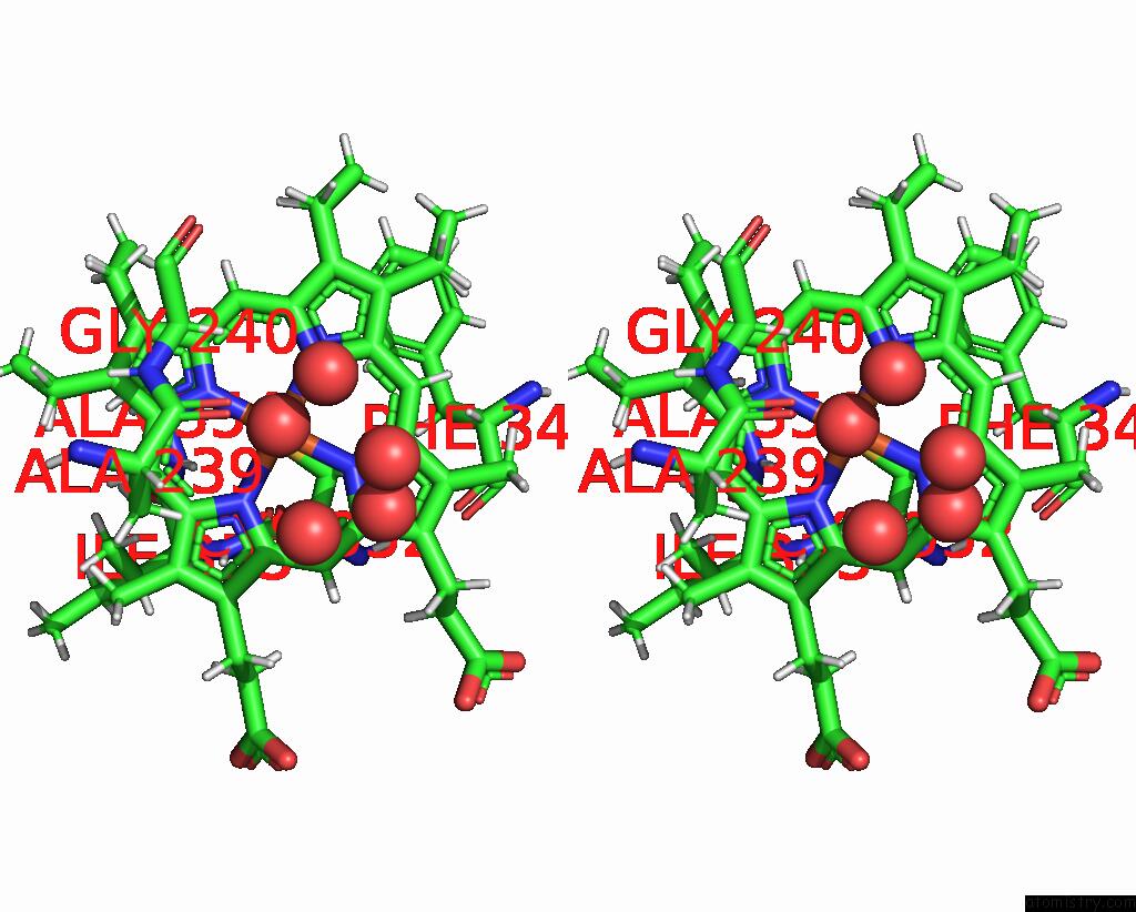
Stereo pair view
Mono view
Stereo pair view
A full contact list of Iron with other atoms in the Fe binding
site number 1 of X-Ray Structure of Nitric Oxide Reductase (Cytochrome P450NOR) in the Ferric Resting State at Atomic Resolution within 5.0Å range:
|
Iron binding site 2 out of 2 in 1jfb
Go back to
Iron binding site 2 out
of 2 in the X-Ray Structure of Nitric Oxide Reductase (Cytochrome P450NOR) in the Ferric Resting State at Atomic Resolution
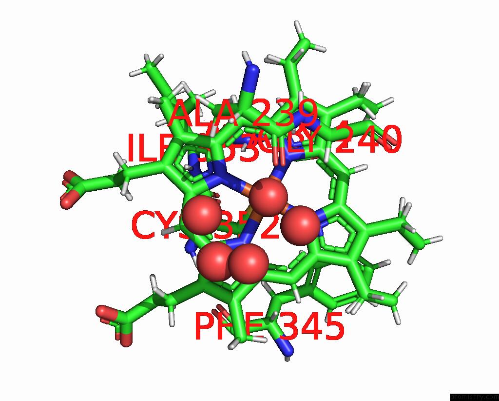
Mono view

Stereo pair view
Mono view
Stereo pair view
A full contact list of Iron with other atoms in the Fe binding
site number 2 of X-Ray Structure of Nitric Oxide Reductase (Cytochrome P450NOR) in the Ferric Resting State at Atomic Resolution within 5.0Å range:
|
Reference:
H.Shimizu,
S.Y.Park,
Y.Shiro,
S.Adachi.
X-Ray Structure of Nitric Oxide Reductase (Cytochrome P450NOR) at Atomic Resolution. Acta Crystallogr.,Sect.D V. 58 81 2002.
ISSN: ISSN 0907-4449
PubMed: 11752781
DOI: 10.1107/S0907444901017383
Page generated: Wed Jul 16 16:31:47 2025
ISSN: ISSN 0907-4449
PubMed: 11752781
DOI: 10.1107/S0907444901017383
Last articles
Fe in 2YXOFe in 2YRS
Fe in 2YXC
Fe in 2YNM
Fe in 2YVJ
Fe in 2YP1
Fe in 2YU2
Fe in 2YU1
Fe in 2YQB
Fe in 2YOO