Iron »
PDB 1j3z-1jju »
1jig »
Iron in PDB 1jig: Dlp-2 From Bacillus Anthracis
Protein crystallography data
The structure of Dlp-2 From Bacillus Anthracis, PDB code: 1jig
was solved by
E.Papinutto,
W.G.Dundon,
N.Pitulis,
R.Battistutta,
C.Montecucco,
G.Zanotti,
with X-Ray Crystallography technique. A brief refinement statistics is given in the table below:
| Resolution Low / High (Å) | 71.63 / 1.46 |
| Space group | H 3 |
| Cell size a, b, c (Å), α, β, γ (°) | 87.723, 87.723, 214.875, 90.00, 90.00, 120.00 |
| R / Rfree (%) | 18.6 / 20.6 |
Iron Binding Sites:
The binding sites of Iron atom in the Dlp-2 From Bacillus Anthracis
(pdb code 1jig). This binding sites where shown within
5.0 Angstroms radius around Iron atom.
In total 4 binding sites of Iron where determined in the Dlp-2 From Bacillus Anthracis, PDB code: 1jig:
Jump to Iron binding site number: 1; 2; 3; 4;
In total 4 binding sites of Iron where determined in the Dlp-2 From Bacillus Anthracis, PDB code: 1jig:
Jump to Iron binding site number: 1; 2; 3; 4;
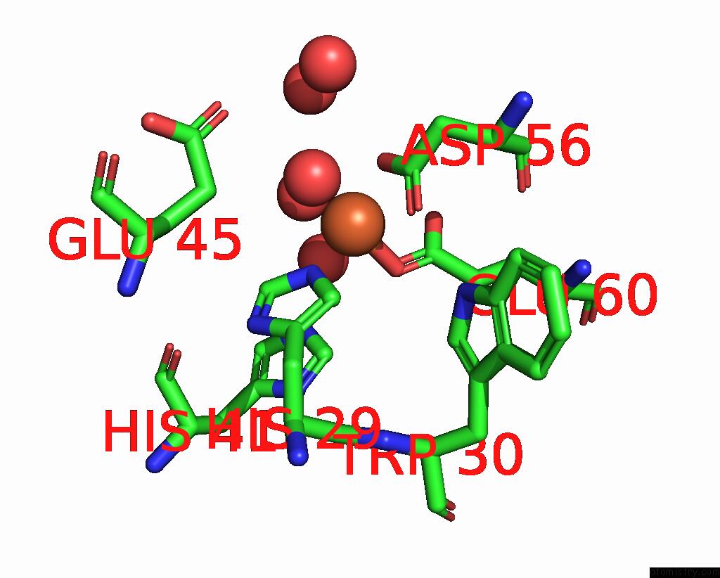
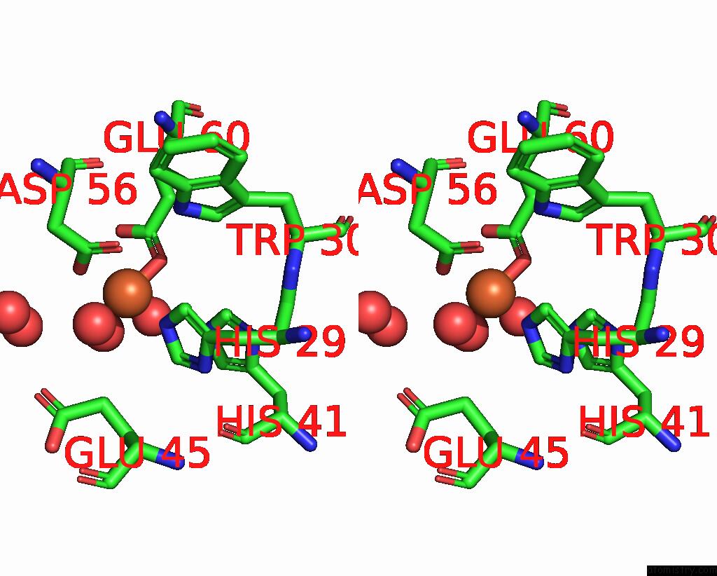
Iron binding site 1 out of 4 in 1jig
Go back to
Iron binding site 1 out
of 4 in the Dlp-2 From Bacillus Anthracis
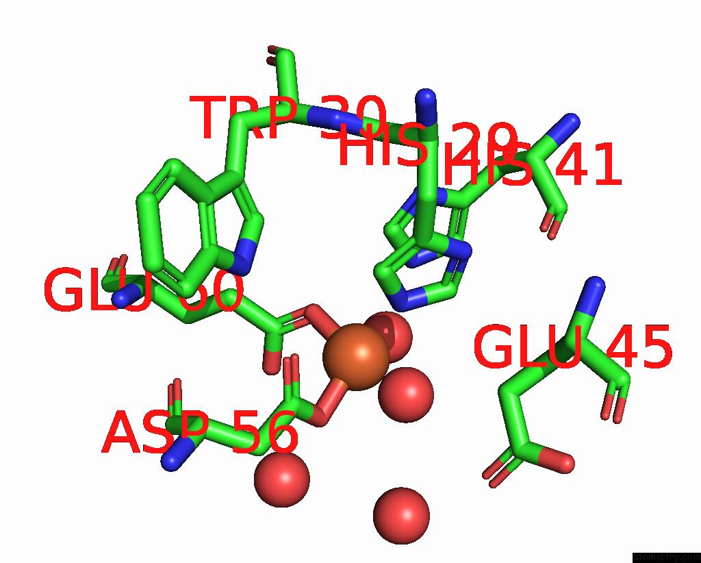
Mono view
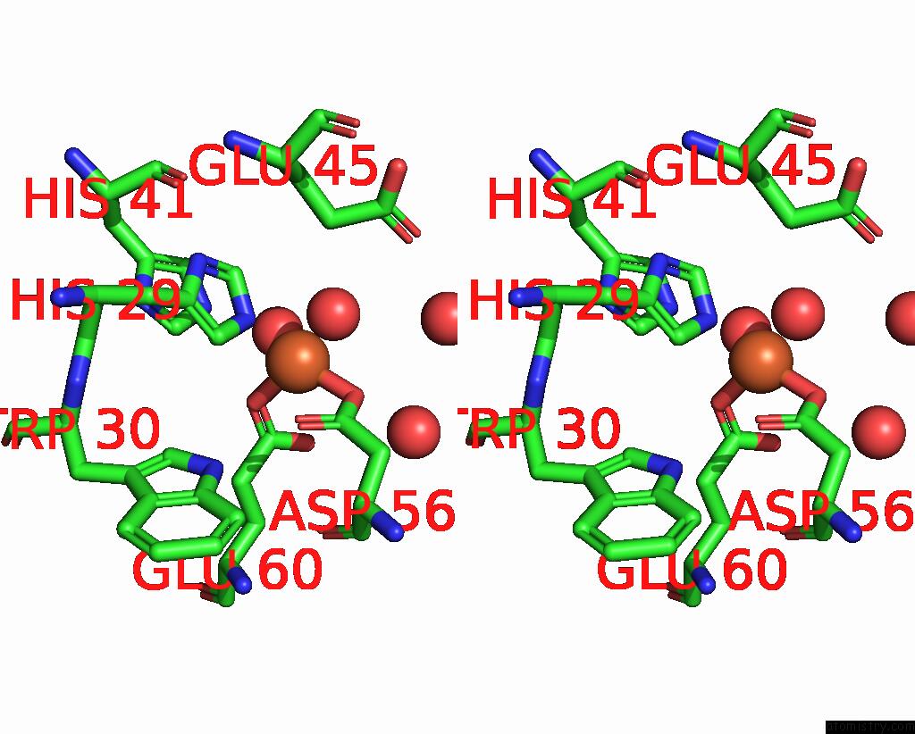
Stereo pair view
Mono view
Stereo pair view
A full contact list of Iron with other atoms in the Fe binding
site number 1 of Dlp-2 From Bacillus Anthracis within 5.0Å range:
|
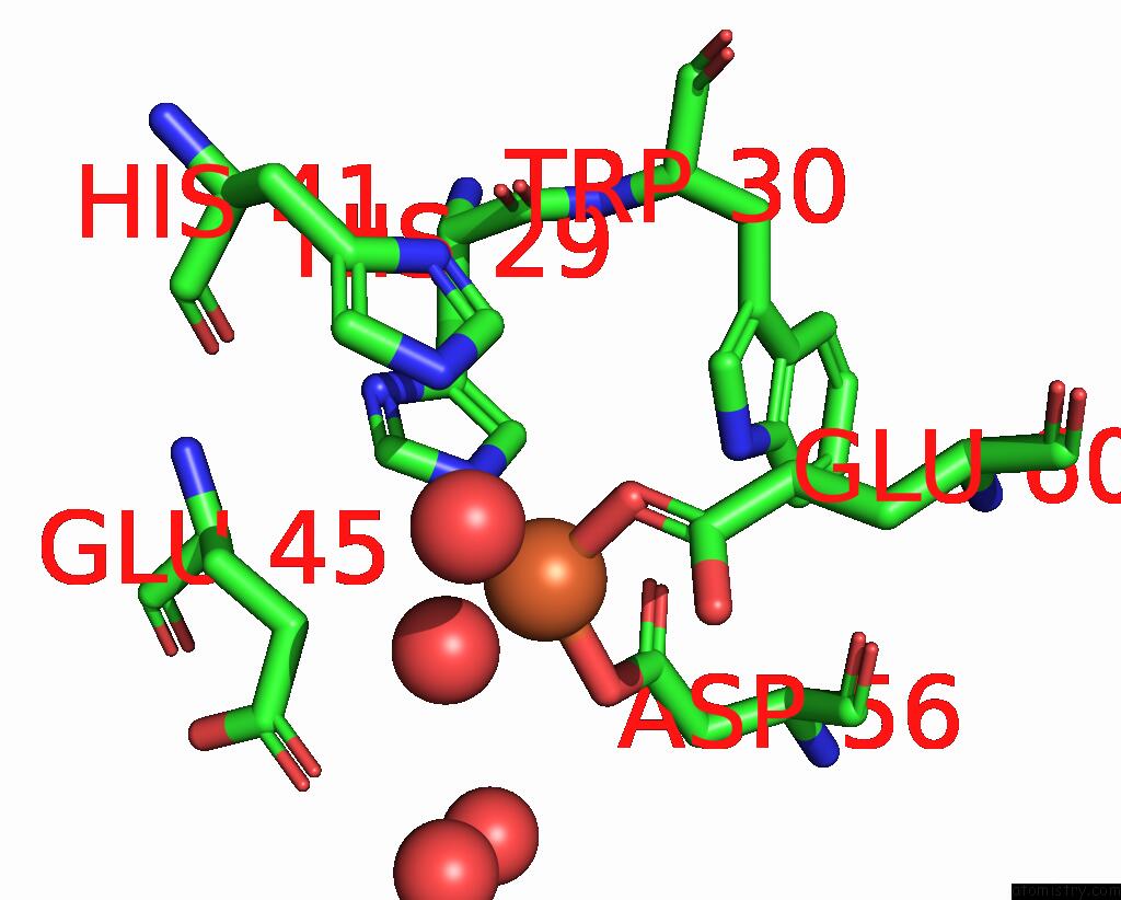
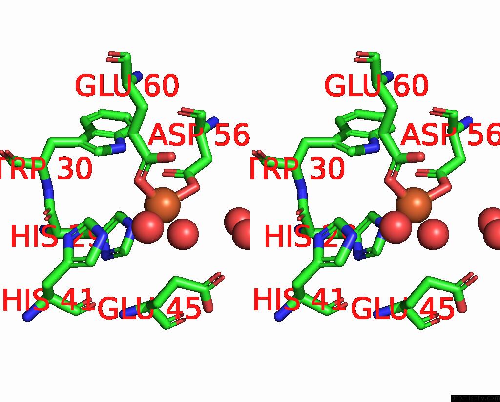
Iron binding site 2 out of 4 in 1jig
Go back to
Iron binding site 2 out
of 4 in the Dlp-2 From Bacillus Anthracis
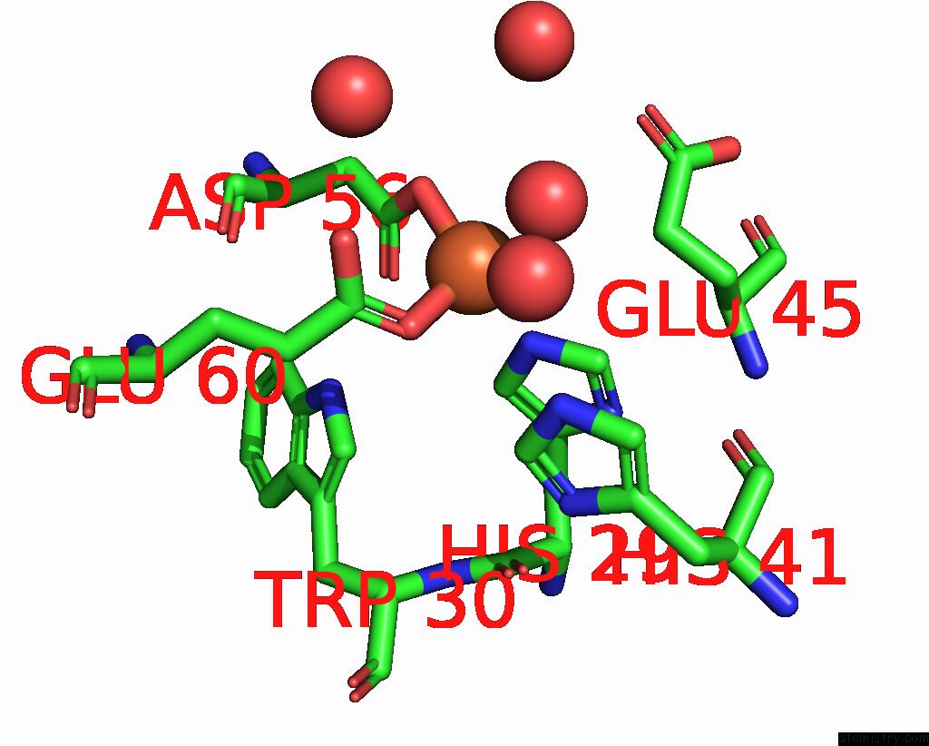
Mono view
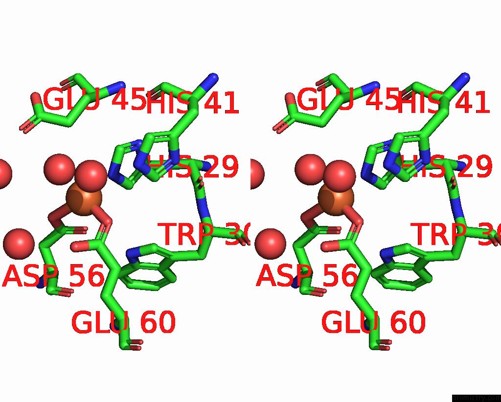
Stereo pair view
Mono view
Stereo pair view
A full contact list of Iron with other atoms in the Fe binding
site number 2 of Dlp-2 From Bacillus Anthracis within 5.0Å range:
|
Iron binding site 3 out of 4 in 1jig
Go back to
Iron binding site 3 out
of 4 in the Dlp-2 From Bacillus Anthracis
Mono view
Stereo pair view
Mono view
Stereo pair view
A full contact list of Iron with other atoms in the Fe binding
site number 3 of Dlp-2 From Bacillus Anthracis within 5.0Å range:
|
Iron binding site 4 out of 4 in 1jig
Go back to
Iron binding site 4 out
of 4 in the Dlp-2 From Bacillus Anthracis
Mono view
Stereo pair view
Mono view
Stereo pair view
A full contact list of Iron with other atoms in the Fe binding
site number 4 of Dlp-2 From Bacillus Anthracis within 5.0Å range:
|
Reference:
E.Papinutto,
W.G.Dundon,
N.Pitulis,
R.Battistutta,
C.Montecucco,
G.Zanotti.
Structure of Two Iron-Binding Proteins From Bacillus Anthracis. J.Biol.Chem. V. 277 15093 2002.
ISSN: ISSN 0021-9258
PubMed: 11836250
DOI: 10.1074/JBC.M112378200
Page generated: Wed Jul 16 16:33:38 2025
ISSN: ISSN 0021-9258
PubMed: 11836250
DOI: 10.1074/JBC.M112378200
Last articles
Fe in 2YXOFe in 2YRS
Fe in 2YXC
Fe in 2YNM
Fe in 2YVJ
Fe in 2YP1
Fe in 2YU2
Fe in 2YU1
Fe in 2YQB
Fe in 2YOO