Iron »
PDB 1j3z-1jju »
1jju »
Iron in PDB 1jju: Structure of A Quinohemoprotein Amine Dehydrogenase with A Unique Redox Cofactor and Highly Unusual Crosslinking
Protein crystallography data
The structure of Structure of A Quinohemoprotein Amine Dehydrogenase with A Unique Redox Cofactor and Highly Unusual Crosslinking, PDB code: 1jju
was solved by
S.Datta,
Y.Mori,
K.Takagi,
K.Kawaguchi,
Z.-W.Chen,
K.Kano,
T.Ikeda,
T.Okajima,
S.Kuroda,
K.Tanizawa,
F.S.Mathews,
with X-Ray Crystallography technique. A brief refinement statistics is given in the table below:
| Resolution Low / High (Å) | 30.00 / 2.05 |
| Space group | P 41 21 2 |
| Cell size a, b, c (Å), α, β, γ (°) | 99.489, 99.489, 214.489, 90.00, 90.00, 90.00 |
| R / Rfree (%) | 20.2 / 25.1 |
Other elements in 1jju:
The structure of Structure of A Quinohemoprotein Amine Dehydrogenase with A Unique Redox Cofactor and Highly Unusual Crosslinking also contains other interesting chemical elements:
| Sodium | (Na) | 1 atom |
Iron Binding Sites:
The binding sites of Iron atom in the Structure of A Quinohemoprotein Amine Dehydrogenase with A Unique Redox Cofactor and Highly Unusual Crosslinking
(pdb code 1jju). This binding sites where shown within
5.0 Angstroms radius around Iron atom.
In total 2 binding sites of Iron where determined in the Structure of A Quinohemoprotein Amine Dehydrogenase with A Unique Redox Cofactor and Highly Unusual Crosslinking, PDB code: 1jju:
Jump to Iron binding site number: 1; 2;
In total 2 binding sites of Iron where determined in the Structure of A Quinohemoprotein Amine Dehydrogenase with A Unique Redox Cofactor and Highly Unusual Crosslinking, PDB code: 1jju:
Jump to Iron binding site number: 1; 2;
Iron binding site 1 out of 2 in 1jju
Go back to
Iron binding site 1 out
of 2 in the Structure of A Quinohemoprotein Amine Dehydrogenase with A Unique Redox Cofactor and Highly Unusual Crosslinking
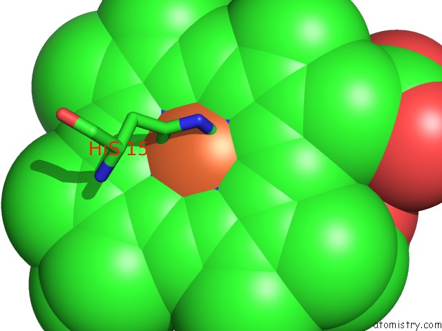
Mono view
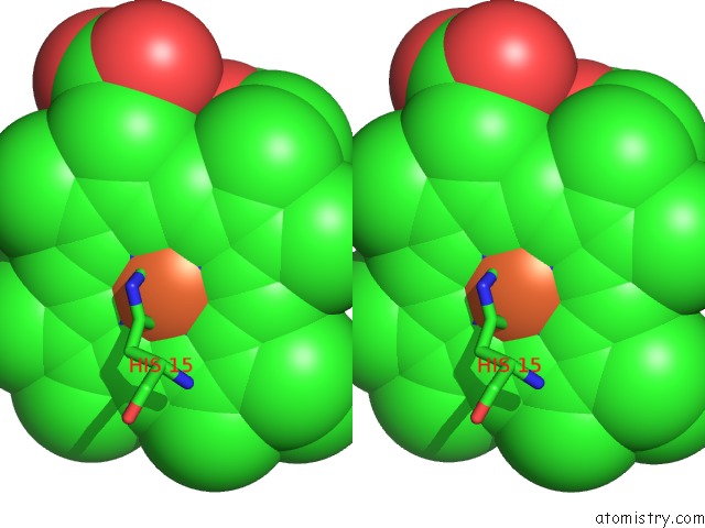
Stereo pair view
Mono view
Stereo pair view
A full contact list of Iron with other atoms in the Fe binding
site number 1 of Structure of A Quinohemoprotein Amine Dehydrogenase with A Unique Redox Cofactor and Highly Unusual Crosslinking within 5.0Å range:
|
Iron binding site 2 out of 2 in 1jju
Go back to
Iron binding site 2 out
of 2 in the Structure of A Quinohemoprotein Amine Dehydrogenase with A Unique Redox Cofactor and Highly Unusual Crosslinking
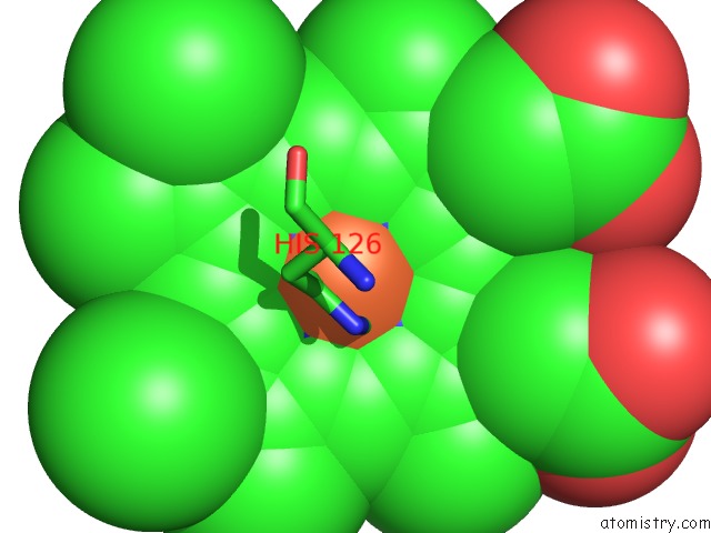
Mono view
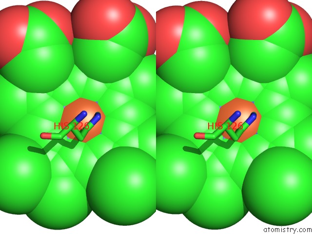
Stereo pair view
Mono view
Stereo pair view
A full contact list of Iron with other atoms in the Fe binding
site number 2 of Structure of A Quinohemoprotein Amine Dehydrogenase with A Unique Redox Cofactor and Highly Unusual Crosslinking within 5.0Å range:
|
Reference:
S.Datta,
Y.Mori,
K.Takagi,
K.Kawaguchi,
Z.W.Chen,
T.Okajima,
S.Kuroda,
T.Ikeda,
K.Kano,
K.Tanizawa,
F.S.Mathews.
Structure of A Quinohemoprotein Amine Dehydrogenase with An Uncommon Redox Cofactor and Highly Unusual Crosslinking. Proc.Natl.Acad.Sci.Usa V. 98 14268 2001.
ISSN: ISSN 0027-8424
PubMed: 11717396
DOI: 10.1073/PNAS.241429098
Page generated: Sat Aug 3 08:32:02 2024
ISSN: ISSN 0027-8424
PubMed: 11717396
DOI: 10.1073/PNAS.241429098
Last articles
Cl in 2Y5TCl in 2Y5H
Cl in 2Y5F
Cl in 2Y5G
Cl in 2Y56
Cl in 2Y58
Cl in 2Y54
Cl in 2Y57
Cl in 2Y4G
Cl in 2Y4R