Iron »
PDB 1jl6-1k2o »
1jqk »
Iron in PDB 1jqk: Crystal Structure of Carbon Monoxide Dehydrogenase From Rhodospirillum Rubrum
Enzymatic activity of Crystal Structure of Carbon Monoxide Dehydrogenase From Rhodospirillum Rubrum
All present enzymatic activity of Crystal Structure of Carbon Monoxide Dehydrogenase From Rhodospirillum Rubrum:
1.2.99.2;
1.2.99.2;
Protein crystallography data
The structure of Crystal Structure of Carbon Monoxide Dehydrogenase From Rhodospirillum Rubrum, PDB code: 1jqk
was solved by
C.L.Drennan,
J.Heo,
M.D.Sintchak,
E.Schreiter,
P.W.Ludden,
with X-Ray Crystallography technique. A brief refinement statistics is given in the table below:
| Resolution Low / High (Å) | 100.00 / 2.80 |
| Space group | P 1 21 1 |
| Cell size a, b, c (Å), α, β, γ (°) | 92.600, 200.100, 116.800, 90.00, 111.50, 90.00 |
| R / Rfree (%) | 25.7 / 28.7 |
Other elements in 1jqk:
The structure of Crystal Structure of Carbon Monoxide Dehydrogenase From Rhodospirillum Rubrum also contains other interesting chemical elements:
| Nickel | (Ni) | 6 atoms |
Iron Binding Sites:
Pages:
>>> Page 1 <<< Page 2, Binding sites: 11 - 20; Page 3, Binding sites: 21 - 30; Page 4, Binding sites: 31 - 40; Page 5, Binding sites: 41 - 50; Page 6, Binding sites: 51 - 60;Binding sites:
The binding sites of Iron atom in the Crystal Structure of Carbon Monoxide Dehydrogenase From Rhodospirillum Rubrum (pdb code 1jqk). This binding sites where shown within 5.0 Angstroms radius around Iron atom.In total 60 binding sites of Iron where determined in the Crystal Structure of Carbon Monoxide Dehydrogenase From Rhodospirillum Rubrum, PDB code: 1jqk:
Jump to Iron binding site number: 1; 2; 3; 4; 5; 6; 7; 8; 9; 10;
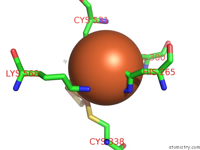
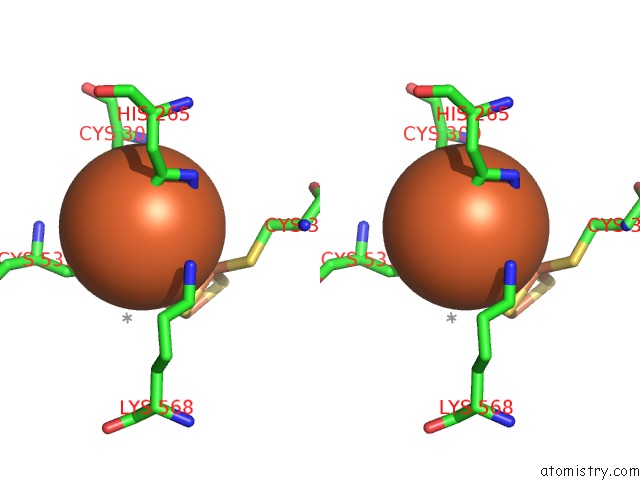
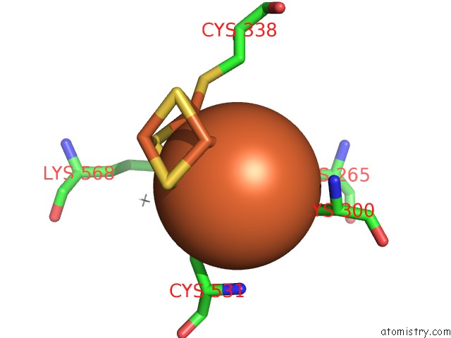
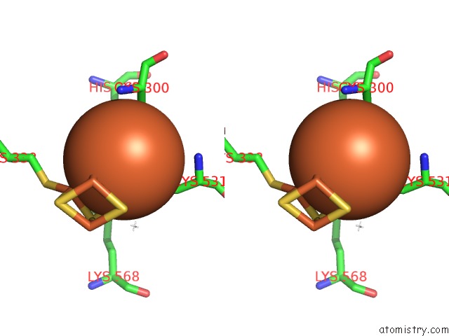
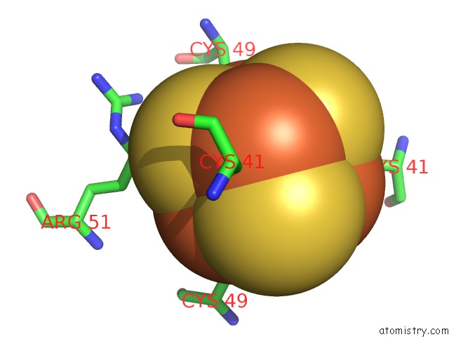
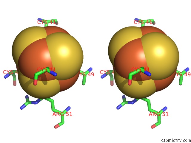
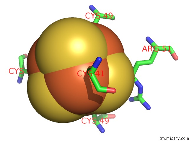
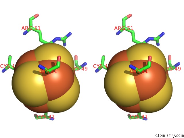
Iron binding site 1 out of 60 in 1jqk
Go back to
Iron binding site 1 out
of 60 in the Crystal Structure of Carbon Monoxide Dehydrogenase From Rhodospirillum Rubrum
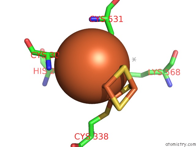
Mono view
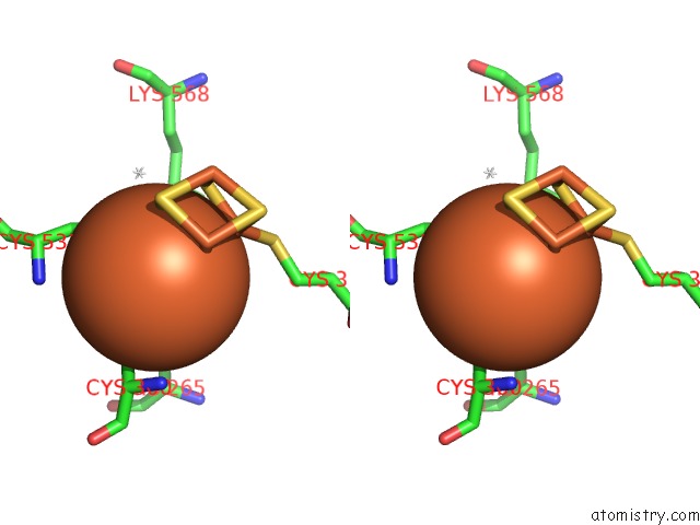
Stereo pair view
Mono view
Stereo pair view
A full contact list of Iron with other atoms in the Fe binding
site number 1 of Crystal Structure of Carbon Monoxide Dehydrogenase From Rhodospirillum Rubrum within 5.0Å range:
|
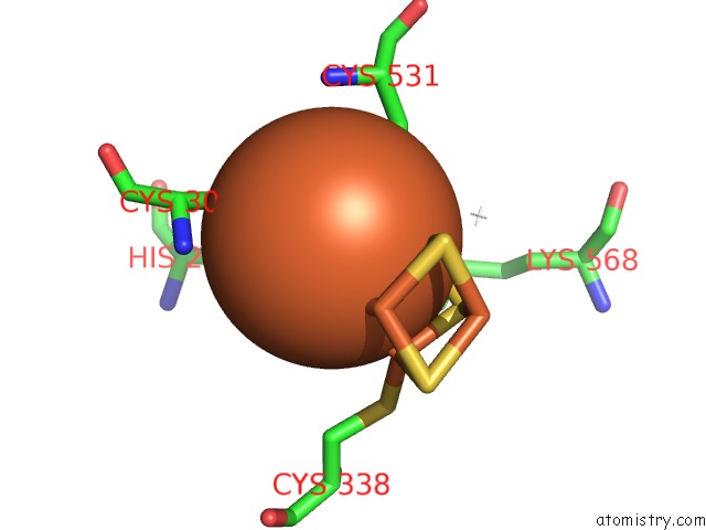
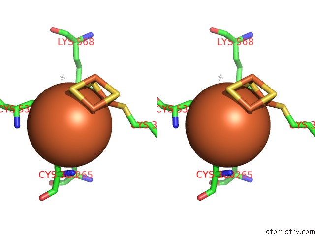
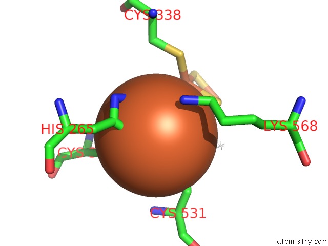
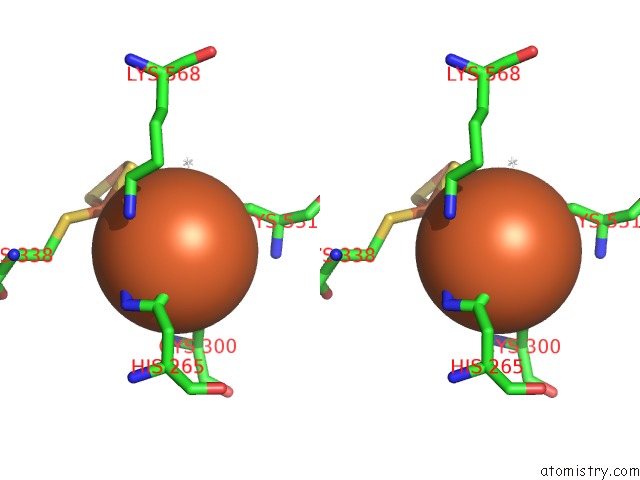
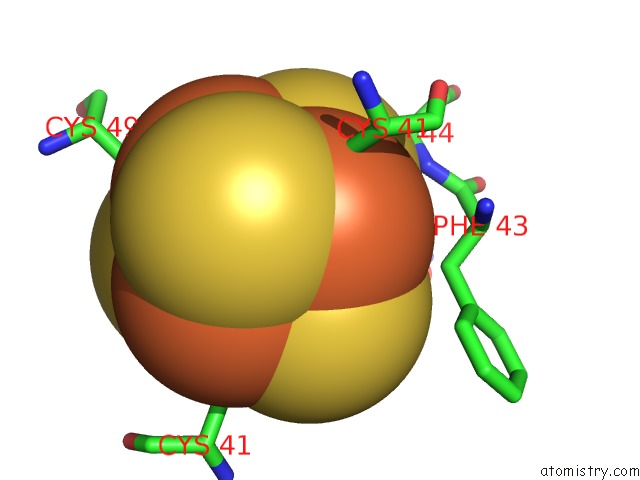
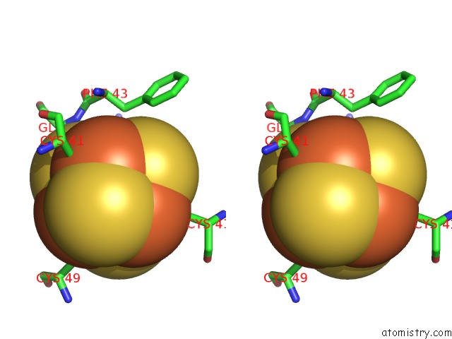
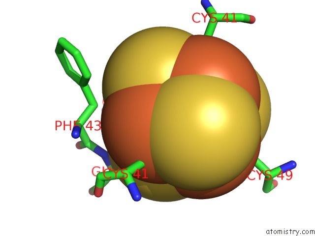
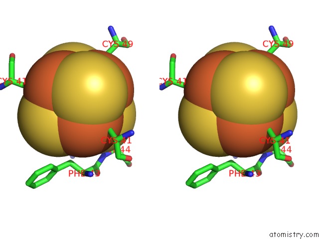
Iron binding site 2 out of 60 in 1jqk
Go back to
Iron binding site 2 out
of 60 in the Crystal Structure of Carbon Monoxide Dehydrogenase From Rhodospirillum Rubrum
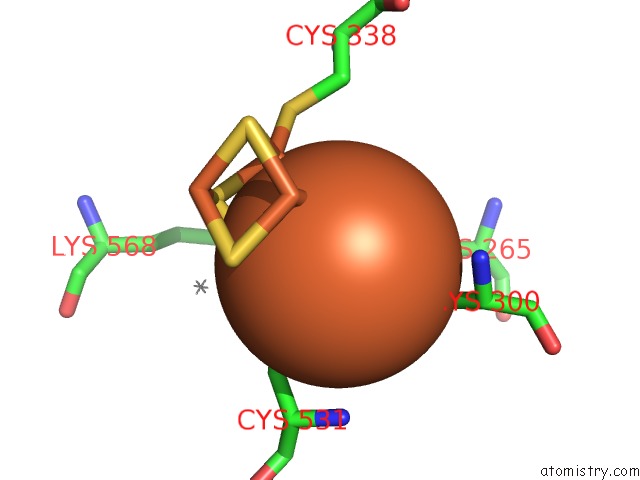
Mono view
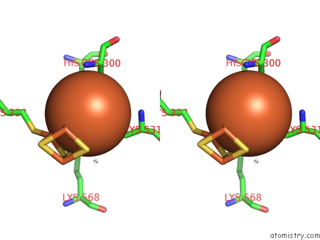
Stereo pair view
Mono view
Stereo pair view
A full contact list of Iron with other atoms in the Fe binding
site number 2 of Crystal Structure of Carbon Monoxide Dehydrogenase From Rhodospirillum Rubrum within 5.0Å range:
|
Iron binding site 3 out of 60 in 1jqk
Go back to
Iron binding site 3 out
of 60 in the Crystal Structure of Carbon Monoxide Dehydrogenase From Rhodospirillum Rubrum
Mono view
Stereo pair view
Mono view
Stereo pair view
A full contact list of Iron with other atoms in the Fe binding
site number 3 of Crystal Structure of Carbon Monoxide Dehydrogenase From Rhodospirillum Rubrum within 5.0Å range:
|
Iron binding site 4 out of 60 in 1jqk
Go back to
Iron binding site 4 out
of 60 in the Crystal Structure of Carbon Monoxide Dehydrogenase From Rhodospirillum Rubrum
Mono view
Stereo pair view
Mono view
Stereo pair view
A full contact list of Iron with other atoms in the Fe binding
site number 4 of Crystal Structure of Carbon Monoxide Dehydrogenase From Rhodospirillum Rubrum within 5.0Å range:
|
Iron binding site 5 out of 60 in 1jqk
Go back to
Iron binding site 5 out
of 60 in the Crystal Structure of Carbon Monoxide Dehydrogenase From Rhodospirillum Rubrum
Mono view
Stereo pair view
Mono view
Stereo pair view
A full contact list of Iron with other atoms in the Fe binding
site number 5 of Crystal Structure of Carbon Monoxide Dehydrogenase From Rhodospirillum Rubrum within 5.0Å range:
|
Iron binding site 6 out of 60 in 1jqk
Go back to
Iron binding site 6 out
of 60 in the Crystal Structure of Carbon Monoxide Dehydrogenase From Rhodospirillum Rubrum
Mono view
Stereo pair view
Mono view
Stereo pair view
A full contact list of Iron with other atoms in the Fe binding
site number 6 of Crystal Structure of Carbon Monoxide Dehydrogenase From Rhodospirillum Rubrum within 5.0Å range:
|
Iron binding site 7 out of 60 in 1jqk
Go back to
Iron binding site 7 out
of 60 in the Crystal Structure of Carbon Monoxide Dehydrogenase From Rhodospirillum Rubrum
Mono view
Stereo pair view
Mono view
Stereo pair view
A full contact list of Iron with other atoms in the Fe binding
site number 7 of Crystal Structure of Carbon Monoxide Dehydrogenase From Rhodospirillum Rubrum within 5.0Å range:
|
Iron binding site 8 out of 60 in 1jqk
Go back to
Iron binding site 8 out
of 60 in the Crystal Structure of Carbon Monoxide Dehydrogenase From Rhodospirillum Rubrum
Mono view
Stereo pair view
Mono view
Stereo pair view
A full contact list of Iron with other atoms in the Fe binding
site number 8 of Crystal Structure of Carbon Monoxide Dehydrogenase From Rhodospirillum Rubrum within 5.0Å range:
|
Iron binding site 9 out of 60 in 1jqk
Go back to
Iron binding site 9 out
of 60 in the Crystal Structure of Carbon Monoxide Dehydrogenase From Rhodospirillum Rubrum
Mono view
Stereo pair view
Mono view
Stereo pair view
A full contact list of Iron with other atoms in the Fe binding
site number 9 of Crystal Structure of Carbon Monoxide Dehydrogenase From Rhodospirillum Rubrum within 5.0Å range:
|
Iron binding site 10 out of 60 in 1jqk
Go back to
Iron binding site 10 out
of 60 in the Crystal Structure of Carbon Monoxide Dehydrogenase From Rhodospirillum Rubrum
Mono view
Stereo pair view
Mono view
Stereo pair view
A full contact list of Iron with other atoms in the Fe binding
site number 10 of Crystal Structure of Carbon Monoxide Dehydrogenase From Rhodospirillum Rubrum within 5.0Å range:
|
Reference:
C.L.Drennan,
J.Heo,
M.D.Sintchak,
E.Schreiter,
P.W.Ludden.
Life on Carbon Monoxide: X-Ray Structure of Rhodospirillum Rubrum Ni-Fe-S Carbon Monoxide Dehydrogenase. Proc.Natl.Acad.Sci.Usa V. 98 11973 2001.
ISSN: ISSN 0027-8424
PubMed: 11593006
DOI: 10.1073/PNAS.211429998
Page generated: Sat Aug 3 08:38:10 2024
ISSN: ISSN 0027-8424
PubMed: 11593006
DOI: 10.1073/PNAS.211429998
Last articles
Zn in 9MJ5Zn in 9HNW
Zn in 9G0L
Zn in 9FNE
Zn in 9DZN
Zn in 9E0I
Zn in 9D32
Zn in 9DAK
Zn in 8ZXC
Zn in 8ZUF