Iron »
PDB 1jl6-1k2o »
1jrp »
Iron in PDB 1jrp: Crystal Structure of Xanthine Dehydrogenase Inhibited By Alloxanthine From Rhodobacter Capsulatus
Enzymatic activity of Crystal Structure of Xanthine Dehydrogenase Inhibited By Alloxanthine From Rhodobacter Capsulatus
All present enzymatic activity of Crystal Structure of Xanthine Dehydrogenase Inhibited By Alloxanthine From Rhodobacter Capsulatus:
1.1.1.204;
1.1.1.204;
Protein crystallography data
The structure of Crystal Structure of Xanthine Dehydrogenase Inhibited By Alloxanthine From Rhodobacter Capsulatus, PDB code: 1jrp
was solved by
J.J.Truglio,
K.Theis,
S.Leimkuhler,
R.Rappa,
K.V.Rajagopalan,
C.Kisker,
with X-Ray Crystallography technique. A brief refinement statistics is given in the table below:
| Resolution Low / High (Å) | 30.00 / 3.00 |
| Space group | P 1 |
| Cell size a, b, c (Å), α, β, γ (°) | 92.617, 140.728, 157.665, 109.59, 105.84, 101.25 |
| R / Rfree (%) | 19.3 / 24.3 |
Other elements in 1jrp:
The structure of Crystal Structure of Xanthine Dehydrogenase Inhibited By Alloxanthine From Rhodobacter Capsulatus also contains other interesting chemical elements:
| Molybdenum | (Mo) | 4 atoms |
| Calcium | (Ca) | 4 atoms |
Iron Binding Sites:
Pages:
>>> Page 1 <<< Page 2, Binding sites: 11 - 16;Binding sites:
The binding sites of Iron atom in the Crystal Structure of Xanthine Dehydrogenase Inhibited By Alloxanthine From Rhodobacter Capsulatus (pdb code 1jrp). This binding sites where shown within 5.0 Angstroms radius around Iron atom.In total 16 binding sites of Iron where determined in the Crystal Structure of Xanthine Dehydrogenase Inhibited By Alloxanthine From Rhodobacter Capsulatus, PDB code: 1jrp:
Jump to Iron binding site number: 1; 2; 3; 4; 5; 6; 7; 8; 9; 10;
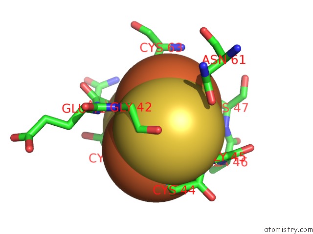
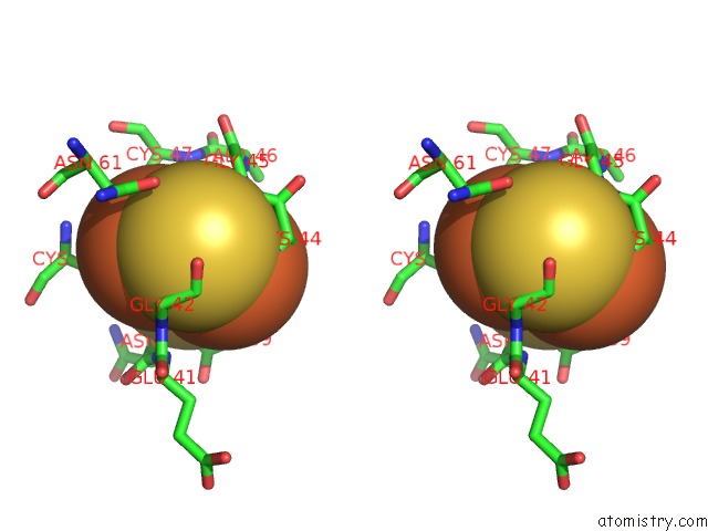
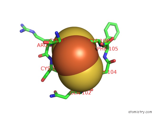
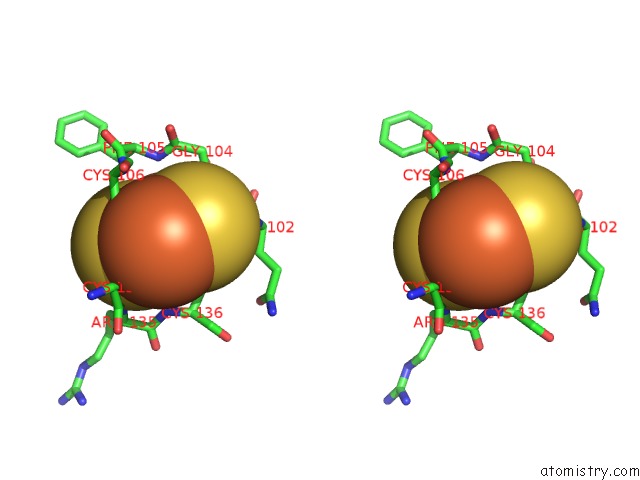
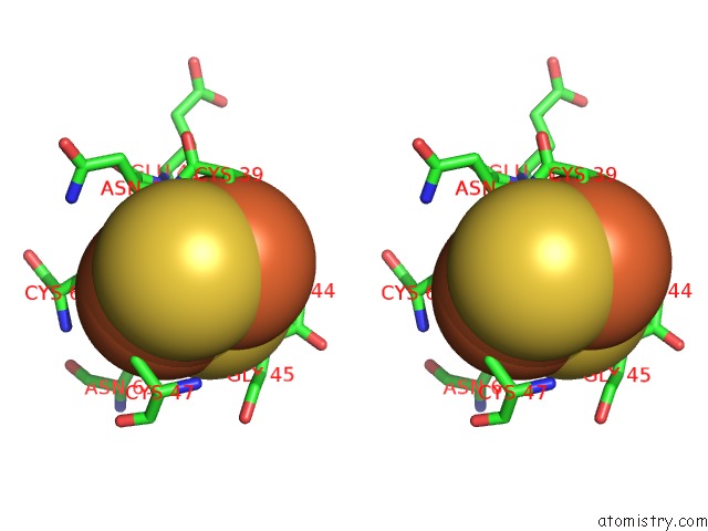
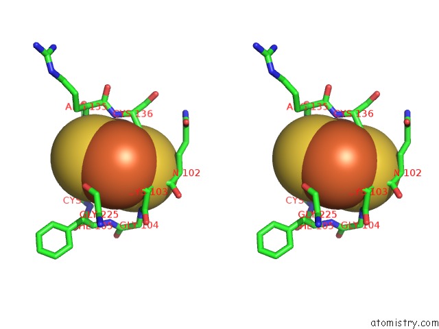
Iron binding site 1 out of 16 in 1jrp
Go back to
Iron binding site 1 out
of 16 in the Crystal Structure of Xanthine Dehydrogenase Inhibited By Alloxanthine From Rhodobacter Capsulatus
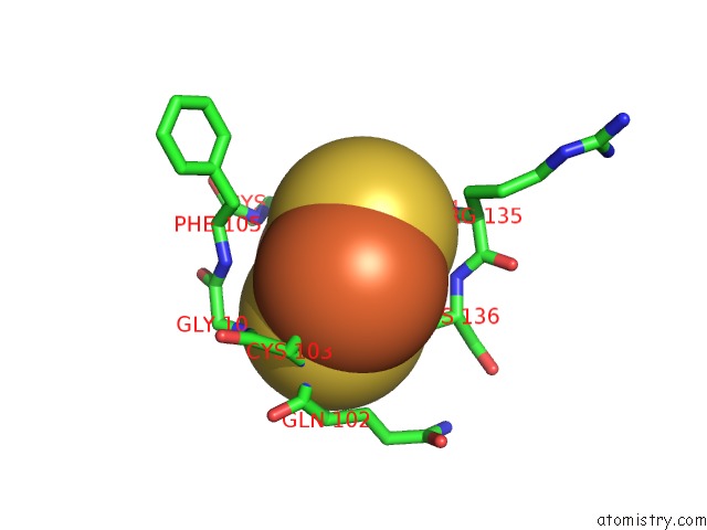
Mono view
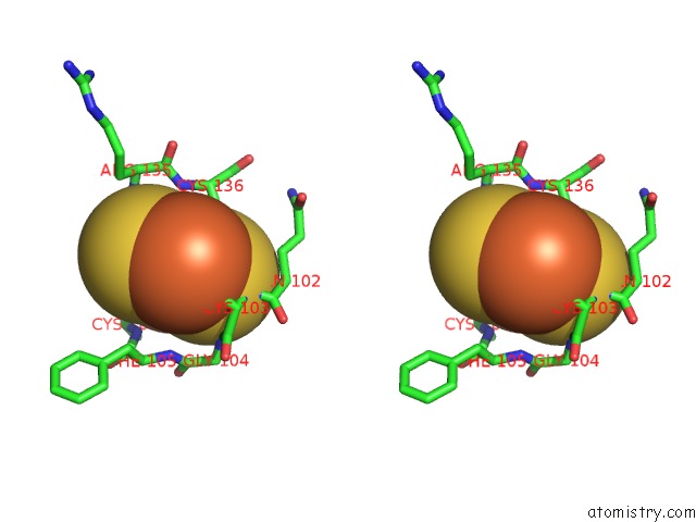
Stereo pair view
Mono view
Stereo pair view
A full contact list of Iron with other atoms in the Fe binding
site number 1 of Crystal Structure of Xanthine Dehydrogenase Inhibited By Alloxanthine From Rhodobacter Capsulatus within 5.0Å range:
|
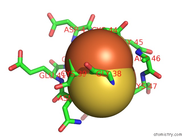
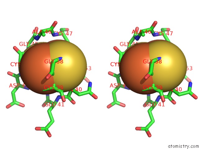
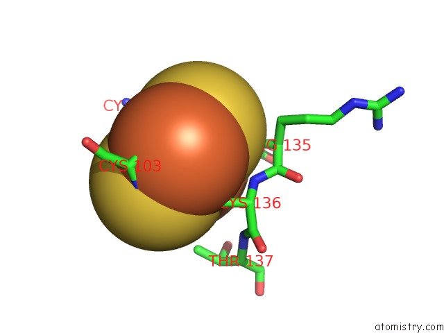
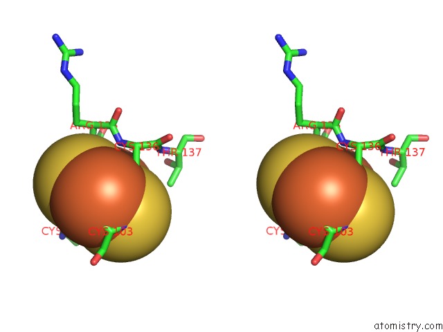
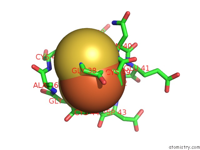
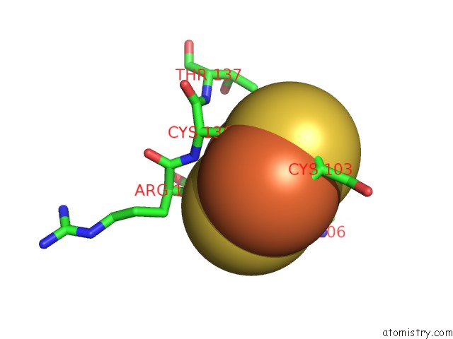
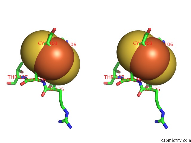
Iron binding site 2 out of 16 in 1jrp
Go back to
Iron binding site 2 out
of 16 in the Crystal Structure of Xanthine Dehydrogenase Inhibited By Alloxanthine From Rhodobacter Capsulatus
Mono view
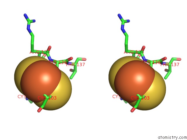
Stereo pair view
Mono view
Stereo pair view
A full contact list of Iron with other atoms in the Fe binding
site number 2 of Crystal Structure of Xanthine Dehydrogenase Inhibited By Alloxanthine From Rhodobacter Capsulatus within 5.0Å range:
|
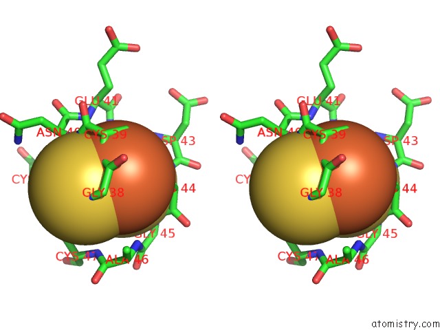
Iron binding site 3 out of 16 in 1jrp
Go back to
Iron binding site 3 out
of 16 in the Crystal Structure of Xanthine Dehydrogenase Inhibited By Alloxanthine From Rhodobacter Capsulatus
Mono view
Stereo pair view
Mono view
Stereo pair view
A full contact list of Iron with other atoms in the Fe binding
site number 3 of Crystal Structure of Xanthine Dehydrogenase Inhibited By Alloxanthine From Rhodobacter Capsulatus within 5.0Å range:
|
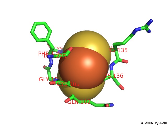
Iron binding site 4 out of 16 in 1jrp
Go back to
Iron binding site 4 out
of 16 in the Crystal Structure of Xanthine Dehydrogenase Inhibited By Alloxanthine From Rhodobacter Capsulatus
Mono view
Stereo pair view
Mono view
Stereo pair view
A full contact list of Iron with other atoms in the Fe binding
site number 4 of Crystal Structure of Xanthine Dehydrogenase Inhibited By Alloxanthine From Rhodobacter Capsulatus within 5.0Å range:
|
Iron binding site 5 out of 16 in 1jrp
Go back to
Iron binding site 5 out
of 16 in the Crystal Structure of Xanthine Dehydrogenase Inhibited By Alloxanthine From Rhodobacter Capsulatus
Mono view
Stereo pair view
Mono view
Stereo pair view
A full contact list of Iron with other atoms in the Fe binding
site number 5 of Crystal Structure of Xanthine Dehydrogenase Inhibited By Alloxanthine From Rhodobacter Capsulatus within 5.0Å range:
|
Iron binding site 6 out of 16 in 1jrp
Go back to
Iron binding site 6 out
of 16 in the Crystal Structure of Xanthine Dehydrogenase Inhibited By Alloxanthine From Rhodobacter Capsulatus
Mono view
Stereo pair view
Mono view
Stereo pair view
A full contact list of Iron with other atoms in the Fe binding
site number 6 of Crystal Structure of Xanthine Dehydrogenase Inhibited By Alloxanthine From Rhodobacter Capsulatus within 5.0Å range:
|
Iron binding site 7 out of 16 in 1jrp
Go back to
Iron binding site 7 out
of 16 in the Crystal Structure of Xanthine Dehydrogenase Inhibited By Alloxanthine From Rhodobacter Capsulatus

Mono view
Stereo pair view
Mono view
Stereo pair view
A full contact list of Iron with other atoms in the Fe binding
site number 7 of Crystal Structure of Xanthine Dehydrogenase Inhibited By Alloxanthine From Rhodobacter Capsulatus within 5.0Å range:
|
Iron binding site 8 out of 16 in 1jrp
Go back to
Iron binding site 8 out
of 16 in the Crystal Structure of Xanthine Dehydrogenase Inhibited By Alloxanthine From Rhodobacter Capsulatus
Mono view
Stereo pair view
Mono view
Stereo pair view
A full contact list of Iron with other atoms in the Fe binding
site number 8 of Crystal Structure of Xanthine Dehydrogenase Inhibited By Alloxanthine From Rhodobacter Capsulatus within 5.0Å range:
|
Iron binding site 9 out of 16 in 1jrp
Go back to
Iron binding site 9 out
of 16 in the Crystal Structure of Xanthine Dehydrogenase Inhibited By Alloxanthine From Rhodobacter Capsulatus
Mono view
Stereo pair view
Mono view
Stereo pair view
A full contact list of Iron with other atoms in the Fe binding
site number 9 of Crystal Structure of Xanthine Dehydrogenase Inhibited By Alloxanthine From Rhodobacter Capsulatus within 5.0Å range:
|
Iron binding site 10 out of 16 in 1jrp
Go back to
Iron binding site 10 out
of 16 in the Crystal Structure of Xanthine Dehydrogenase Inhibited By Alloxanthine From Rhodobacter Capsulatus
Mono view
Stereo pair view
Mono view
Stereo pair view
A full contact list of Iron with other atoms in the Fe binding
site number 10 of Crystal Structure of Xanthine Dehydrogenase Inhibited By Alloxanthine From Rhodobacter Capsulatus within 5.0Å range:
|
Reference:
J.J.Truglio,
K.Theis,
S.Leimkuhler,
R.Rappa,
K.V.Rajagopalan,
C.Kisker.
Crystal Structures of the Active and Alloxanthine-Inhibited Forms of Xanthine Dehydrogenase From Rhodobacter Capsulatus Structure V. 10 115 2002.
ISSN: ISSN 0969-2126
PubMed: 11796116
DOI: 10.1016/S0969-2126(01)00697-9
Page generated: Sat Aug 3 08:38:30 2024
ISSN: ISSN 0969-2126
PubMed: 11796116
DOI: 10.1016/S0969-2126(01)00697-9
Last articles
Zn in 9JYWZn in 9IR4
Zn in 9IR3
Zn in 9GMX
Zn in 9GMW
Zn in 9JEJ
Zn in 9ERF
Zn in 9ERE
Zn in 9EGV
Zn in 9EGW