Iron »
PDB 1k2r-1kqg »
1k6w »
Iron in PDB 1k6w: The Structure of Escherichia Coli Cytosine Deaminase
Enzymatic activity of The Structure of Escherichia Coli Cytosine Deaminase
All present enzymatic activity of The Structure of Escherichia Coli Cytosine Deaminase:
3.5.4.1;
3.5.4.1;
Protein crystallography data
The structure of The Structure of Escherichia Coli Cytosine Deaminase, PDB code: 1k6w
was solved by
G.C.Ireton,
G.Mcdermott,
M.E.Black,
B.L.Stoddard,
with X-Ray Crystallography technique. A brief refinement statistics is given in the table below:
| Resolution Low / High (Å) | 19.98 / 1.75 |
| Space group | H 3 2 |
| Cell size a, b, c (Å), α, β, γ (°) | 109.150, 109.150, 240.050, 90.00, 90.00, 120.00 |
| R / Rfree (%) | 15.5 / 17.2 |
Iron Binding Sites:
The binding sites of Iron atom in the The Structure of Escherichia Coli Cytosine Deaminase
(pdb code 1k6w). This binding sites where shown within
5.0 Angstroms radius around Iron atom.
In total only one binding site of Iron was determined in the The Structure of Escherichia Coli Cytosine Deaminase, PDB code: 1k6w:
In total only one binding site of Iron was determined in the The Structure of Escherichia Coli Cytosine Deaminase, PDB code: 1k6w:
Iron binding site 1 out of 1 in 1k6w
Go back to
Iron binding site 1 out
of 1 in the The Structure of Escherichia Coli Cytosine Deaminase
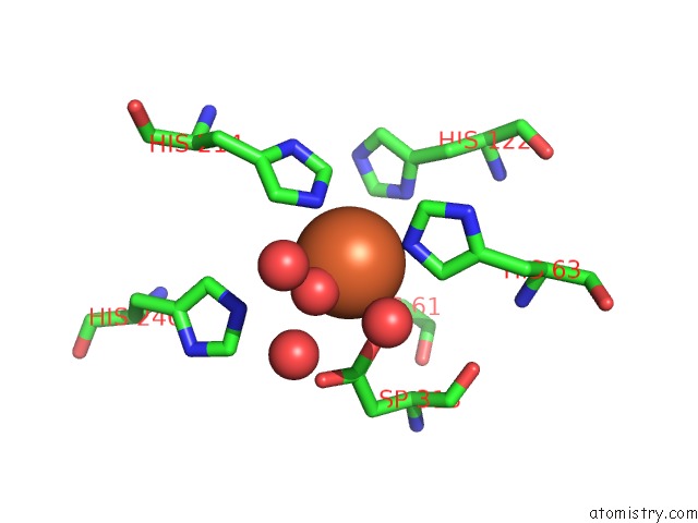
Mono view
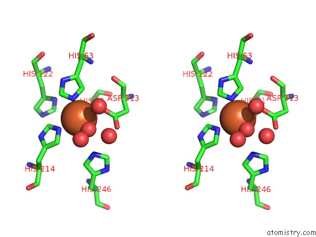
Stereo pair view
Mono view
Stereo pair view
A full contact list of Iron with other atoms in the Fe binding
site number 1 of The Structure of Escherichia Coli Cytosine Deaminase within 5.0Å range:
|
Reference:
G.C.Ireton,
G.Mcdermott,
M.E.Black,
B.L.Stoddard.
The Structure of Escherichia Coli Cytosine Deaminase. J.Mol.Biol. V. 315 687 2002.
ISSN: ISSN 0022-2836
PubMed: 11812140
DOI: 10.1006/JMBI.2001.5277
Page generated: Sat Aug 3 09:06:21 2024
ISSN: ISSN 0022-2836
PubMed: 11812140
DOI: 10.1006/JMBI.2001.5277
Last articles
Cl in 5YJHCl in 5YJG
Cl in 5YKA
Cl in 5YJS
Cl in 5YJP
Cl in 5YJK
Cl in 5YJM
Cl in 5YHO
Cl in 5YIY
Cl in 5YG4