Iron »
PDB 1k2r-1kqg »
1kb0 »
Iron in PDB 1kb0: Crystal Structure of Quinohemoprotein Alcohol Dehydrogenase From Comamonas Testosteroni
Protein crystallography data
The structure of Crystal Structure of Quinohemoprotein Alcohol Dehydrogenase From Comamonas Testosteroni, PDB code: 1kb0
was solved by
H.J.Rozeboom,
A.Oubrie,
with X-Ray Crystallography technique. A brief refinement statistics is given in the table below:
| Resolution Low / High (Å) | 29.63 / 1.44 |
| Space group | C 1 2 1 |
| Cell size a, b, c (Å), α, β, γ (°) | 97.920, 74.331, 92.332, 90.00, 105.67, 90.00 |
| R / Rfree (%) | 16 / 18.8 |
Other elements in 1kb0:
The structure of Crystal Structure of Quinohemoprotein Alcohol Dehydrogenase From Comamonas Testosteroni also contains other interesting chemical elements:
| Calcium | (Ca) | 1 atom |
Iron Binding Sites:
The binding sites of Iron atom in the Crystal Structure of Quinohemoprotein Alcohol Dehydrogenase From Comamonas Testosteroni
(pdb code 1kb0). This binding sites where shown within
5.0 Angstroms radius around Iron atom.
In total only one binding site of Iron was determined in the Crystal Structure of Quinohemoprotein Alcohol Dehydrogenase From Comamonas Testosteroni, PDB code: 1kb0:
In total only one binding site of Iron was determined in the Crystal Structure of Quinohemoprotein Alcohol Dehydrogenase From Comamonas Testosteroni, PDB code: 1kb0:
Iron binding site 1 out of 1 in 1kb0
Go back to
Iron binding site 1 out
of 1 in the Crystal Structure of Quinohemoprotein Alcohol Dehydrogenase From Comamonas Testosteroni
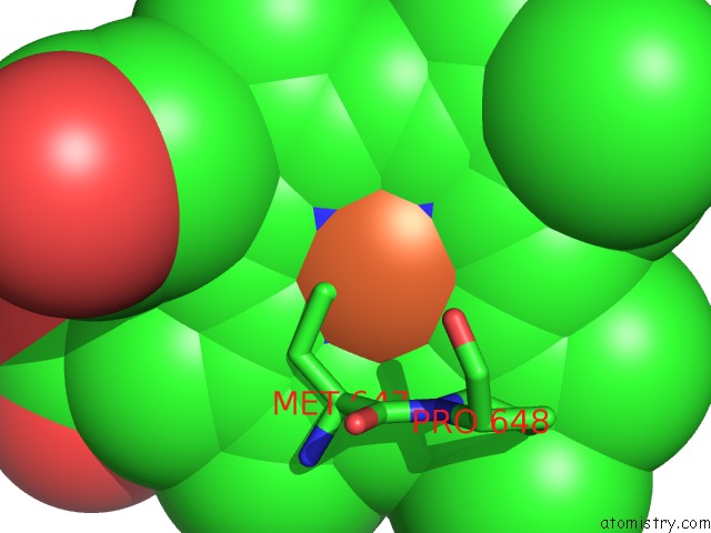
Mono view
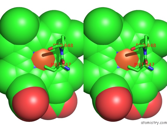
Stereo pair view
Mono view
Stereo pair view
A full contact list of Iron with other atoms in the Fe binding
site number 1 of Crystal Structure of Quinohemoprotein Alcohol Dehydrogenase From Comamonas Testosteroni within 5.0Å range:
|
Reference:
A.Oubrie,
H.J.Rozeboom,
K.H.Kalk,
E.G.Huizinga,
B.W.Dijkstra.
Crystal Structure of Quinohemoprotein Alcohol Dehydrogenase From Comamonas Testosteroni: Structural Basis For Substrate Oxidation and Electron Transfer. J.Biol.Chem. V. 277 3727 2002.
ISSN: ISSN 0021-9258
PubMed: 11714714
DOI: 10.1074/JBC.M109403200
Page generated: Wed Jul 16 16:56:32 2025
ISSN: ISSN 0021-9258
PubMed: 11714714
DOI: 10.1074/JBC.M109403200
Last articles
Fe in 2YXOFe in 2YRS
Fe in 2YXC
Fe in 2YNM
Fe in 2YVJ
Fe in 2YP1
Fe in 2YU2
Fe in 2YU1
Fe in 2YQB
Fe in 2YOO