Iron »
PDB 1k2r-1kqg »
1kg3 »
Iron in PDB 1kg3: Crystal Structure of the Core Fragment of Muty From E.Coli at 1.55A Resolution
Protein crystallography data
The structure of Crystal Structure of the Core Fragment of Muty From E.Coli at 1.55A Resolution, PDB code: 1kg3
was solved by
R.Gilboa,
A.Kilshtein,
D.O.Zharkov,
J.H.Kycia,
S.E.Gerchman,
A.P.Grollman,
G.Shoham,
with X-Ray Crystallography technique. A brief refinement statistics is given in the table below:
| Resolution Low / High (Å) | 43.00 / 1.55 |
| Space group | C 1 2 1 |
| Cell size a, b, c (Å), α, β, γ (°) | 84.450, 50.200, 70.500, 90.00, 123.29, 90.00 |
| R / Rfree (%) | 17 / 22.8 |
Iron Binding Sites:
The binding sites of Iron atom in the Crystal Structure of the Core Fragment of Muty From E.Coli at 1.55A Resolution
(pdb code 1kg3). This binding sites where shown within
5.0 Angstroms radius around Iron atom.
In total 4 binding sites of Iron where determined in the Crystal Structure of the Core Fragment of Muty From E.Coli at 1.55A Resolution, PDB code: 1kg3:
Jump to Iron binding site number: 1; 2; 3; 4;
In total 4 binding sites of Iron where determined in the Crystal Structure of the Core Fragment of Muty From E.Coli at 1.55A Resolution, PDB code: 1kg3:
Jump to Iron binding site number: 1; 2; 3; 4;
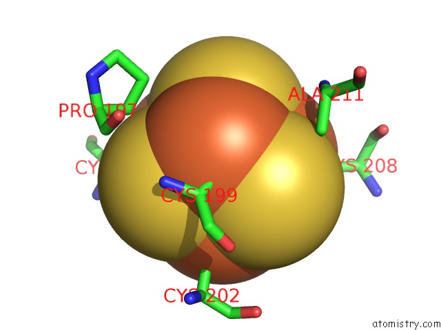
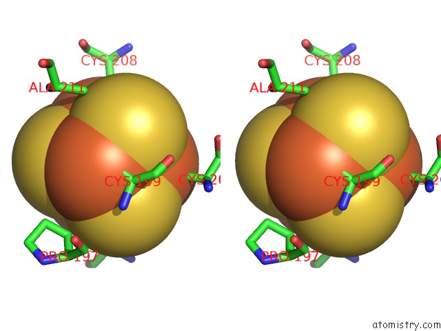
Iron binding site 1 out of 4 in 1kg3
Go back to
Iron binding site 1 out
of 4 in the Crystal Structure of the Core Fragment of Muty From E.Coli at 1.55A Resolution
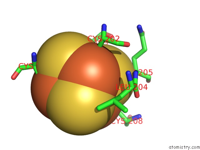
Mono view
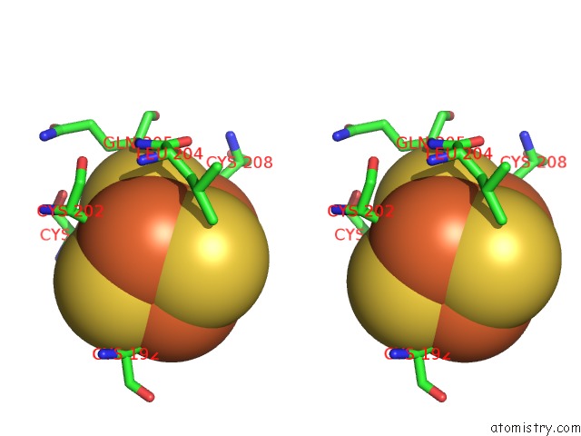
Stereo pair view
Mono view
Stereo pair view
A full contact list of Iron with other atoms in the Fe binding
site number 1 of Crystal Structure of the Core Fragment of Muty From E.Coli at 1.55A Resolution within 5.0Å range:
|
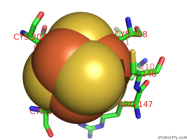
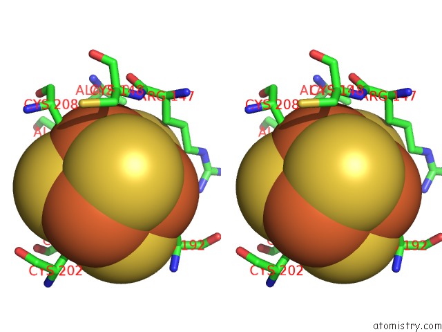
Iron binding site 2 out of 4 in 1kg3
Go back to
Iron binding site 2 out
of 4 in the Crystal Structure of the Core Fragment of Muty From E.Coli at 1.55A Resolution
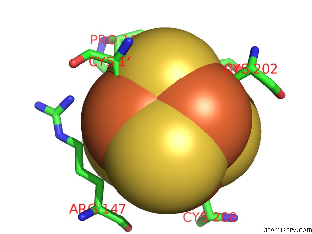
Mono view
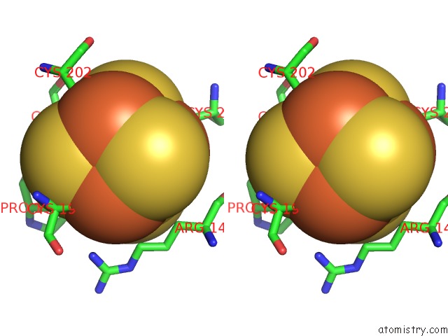
Stereo pair view
Mono view
Stereo pair view
A full contact list of Iron with other atoms in the Fe binding
site number 2 of Crystal Structure of the Core Fragment of Muty From E.Coli at 1.55A Resolution within 5.0Å range:
|
Iron binding site 3 out of 4 in 1kg3
Go back to
Iron binding site 3 out
of 4 in the Crystal Structure of the Core Fragment of Muty From E.Coli at 1.55A Resolution
Mono view
Stereo pair view
Mono view
Stereo pair view
A full contact list of Iron with other atoms in the Fe binding
site number 3 of Crystal Structure of the Core Fragment of Muty From E.Coli at 1.55A Resolution within 5.0Å range:
|
Iron binding site 4 out of 4 in 1kg3
Go back to
Iron binding site 4 out
of 4 in the Crystal Structure of the Core Fragment of Muty From E.Coli at 1.55A Resolution
Mono view
Stereo pair view
Mono view
Stereo pair view
A full contact list of Iron with other atoms in the Fe binding
site number 4 of Crystal Structure of the Core Fragment of Muty From E.Coli at 1.55A Resolution within 5.0Å range:
|
Reference:
R.Gilboa,
A.Kilshtein,
D.O.Zharkov,
J.H.Kycia,
S.E.Gerchman,
A.P.Grollman,
G.Shoham.
Analysis of the E.Coli Muty Dna Glycosylase Structure and Function By Site-Directed Mutagenesis To Be Published.
Page generated: Sat Aug 3 09:09:11 2024
Last articles
Zn in 9MJ5Zn in 9HNW
Zn in 9G0L
Zn in 9FNE
Zn in 9DZN
Zn in 9E0I
Zn in 9D32
Zn in 9DAK
Zn in 8ZXC
Zn in 8ZUF