Iron »
PDB 1k2r-1kqg »
1kg6 »
Iron in PDB 1kg6: Crystal Structure of the K142R Mutant of E.Coli Muty (Core Fragment)
Protein crystallography data
The structure of Crystal Structure of the K142R Mutant of E.Coli Muty (Core Fragment), PDB code: 1kg6
was solved by
R.Gilboa,
A.Kilshtein,
D.O.Zharkov,
J.H.Kycia,
S.E.Gerchman,
A.P.Grollman,
G.Shoham,
with X-Ray Crystallography technique. A brief refinement statistics is given in the table below:
| Resolution Low / High (Å) | 42.00 / 1.50 |
| Space group | C 1 2 1 |
| Cell size a, b, c (Å), α, β, γ (°) | 82.730, 49.950, 69.660, 90.00, 122.74, 90.00 |
| R / Rfree (%) | 15.5 / 19.6 |
Iron Binding Sites:
The binding sites of Iron atom in the Crystal Structure of the K142R Mutant of E.Coli Muty (Core Fragment)
(pdb code 1kg6). This binding sites where shown within
5.0 Angstroms radius around Iron atom.
In total 4 binding sites of Iron where determined in the Crystal Structure of the K142R Mutant of E.Coli Muty (Core Fragment), PDB code: 1kg6:
Jump to Iron binding site number: 1; 2; 3; 4;
In total 4 binding sites of Iron where determined in the Crystal Structure of the K142R Mutant of E.Coli Muty (Core Fragment), PDB code: 1kg6:
Jump to Iron binding site number: 1; 2; 3; 4;
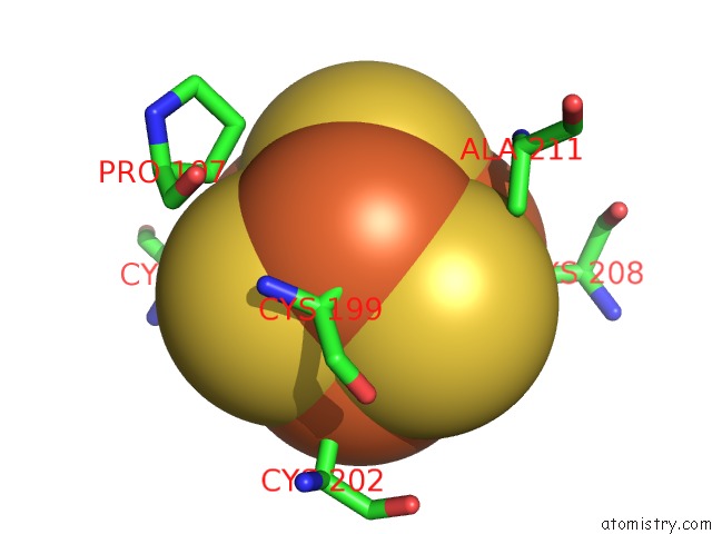
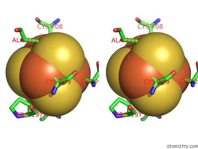
Iron binding site 1 out of 4 in 1kg6
Go back to
Iron binding site 1 out
of 4 in the Crystal Structure of the K142R Mutant of E.Coli Muty (Core Fragment)
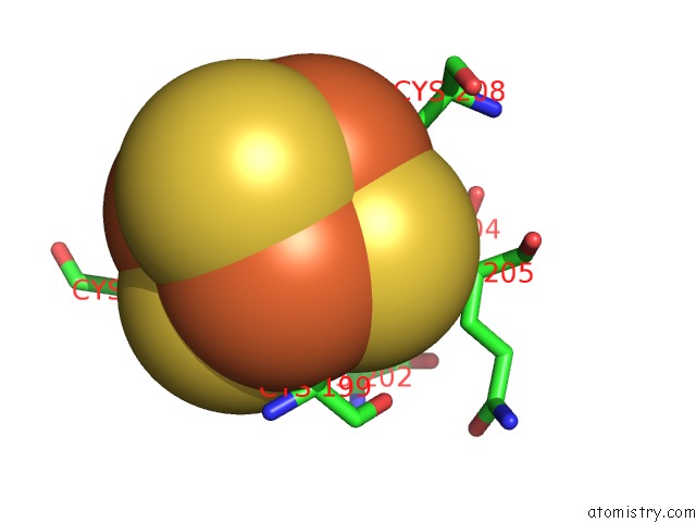
Mono view
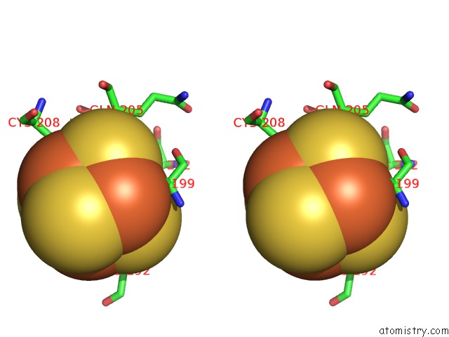
Stereo pair view
Mono view
Stereo pair view
A full contact list of Iron with other atoms in the Fe binding
site number 1 of Crystal Structure of the K142R Mutant of E.Coli Muty (Core Fragment) within 5.0Å range:
|
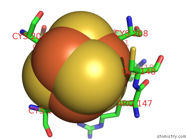
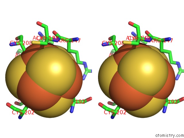
Iron binding site 2 out of 4 in 1kg6
Go back to
Iron binding site 2 out
of 4 in the Crystal Structure of the K142R Mutant of E.Coli Muty (Core Fragment)
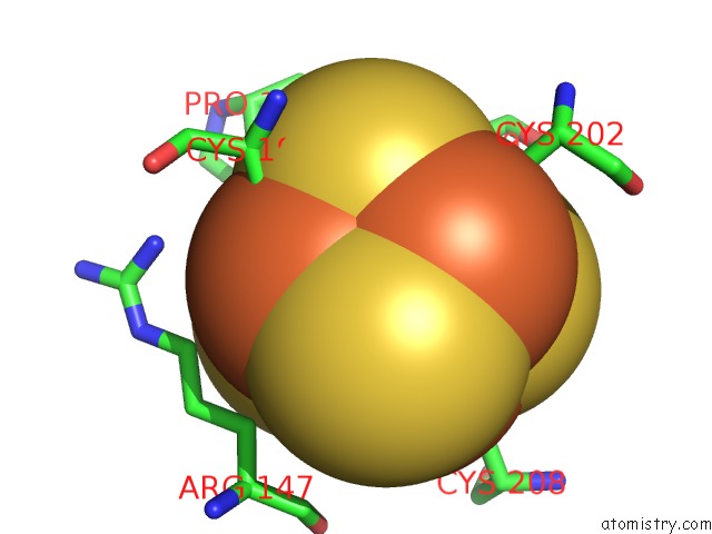
Mono view
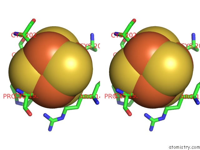
Stereo pair view
Mono view
Stereo pair view
A full contact list of Iron with other atoms in the Fe binding
site number 2 of Crystal Structure of the K142R Mutant of E.Coli Muty (Core Fragment) within 5.0Å range:
|
Iron binding site 3 out of 4 in 1kg6
Go back to
Iron binding site 3 out
of 4 in the Crystal Structure of the K142R Mutant of E.Coli Muty (Core Fragment)
Mono view
Stereo pair view
Mono view
Stereo pair view
A full contact list of Iron with other atoms in the Fe binding
site number 3 of Crystal Structure of the K142R Mutant of E.Coli Muty (Core Fragment) within 5.0Å range:
|
Iron binding site 4 out of 4 in 1kg6
Go back to
Iron binding site 4 out
of 4 in the Crystal Structure of the K142R Mutant of E.Coli Muty (Core Fragment)
Mono view
Stereo pair view
Mono view
Stereo pair view
A full contact list of Iron with other atoms in the Fe binding
site number 4 of Crystal Structure of the K142R Mutant of E.Coli Muty (Core Fragment) within 5.0Å range:
|
Reference:
R.Gilboa,
A.Kilshtein,
D.O.Zharkov,
J.H.Kycia,
S.E.Gerchman,
A.P.Grollman,
G.Shoham.
Analysis of the E.Coli Muty Dna Glycosylase Structure and Function By Site-Directed Mutagenesis To Be Published.
Page generated: Wed Jul 16 17:04:17 2025
Last articles
Fe in 2YXOFe in 2YRS
Fe in 2YXC
Fe in 2YNM
Fe in 2YVJ
Fe in 2YP1
Fe in 2YU2
Fe in 2YU1
Fe in 2YQB
Fe in 2YOO