Iron »
PDB 1lfk-1lr6 »
1lfk »
Iron in PDB 1lfk: Crystal Structure of Oxyb, A Cytochrome P450 Implicated in An Oxidative Phenol Coupling Reaction During Vancomycin Biosynthesis
Protein crystallography data
The structure of Crystal Structure of Oxyb, A Cytochrome P450 Implicated in An Oxidative Phenol Coupling Reaction During Vancomycin Biosynthesis, PDB code: 1lfk
was solved by
O.Pylypenko,
K.Zerbe,
F.Vitali,
W.Zhang,
J.W.Vrijbloed,
J.A.Robinson,
I.Schlichting,
with X-Ray Crystallography technique. A brief refinement statistics is given in the table below:
| Resolution Low / High (Å) | 28.80 / 1.70 |
| Space group | C 1 2 1 |
| Cell size a, b, c (Å), α, β, γ (°) | 100.400, 60.400, 90.300, 90.00, 122.70, 90.00 |
| R / Rfree (%) | 20.3 / 23.5 |
Iron Binding Sites:
The binding sites of Iron atom in the Crystal Structure of Oxyb, A Cytochrome P450 Implicated in An Oxidative Phenol Coupling Reaction During Vancomycin Biosynthesis
(pdb code 1lfk). This binding sites where shown within
5.0 Angstroms radius around Iron atom.
In total only one binding site of Iron was determined in the Crystal Structure of Oxyb, A Cytochrome P450 Implicated in An Oxidative Phenol Coupling Reaction During Vancomycin Biosynthesis, PDB code: 1lfk:
In total only one binding site of Iron was determined in the Crystal Structure of Oxyb, A Cytochrome P450 Implicated in An Oxidative Phenol Coupling Reaction During Vancomycin Biosynthesis, PDB code: 1lfk:
Iron binding site 1 out of 1 in 1lfk
Go back to
Iron binding site 1 out
of 1 in the Crystal Structure of Oxyb, A Cytochrome P450 Implicated in An Oxidative Phenol Coupling Reaction During Vancomycin Biosynthesis
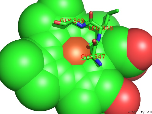
Mono view
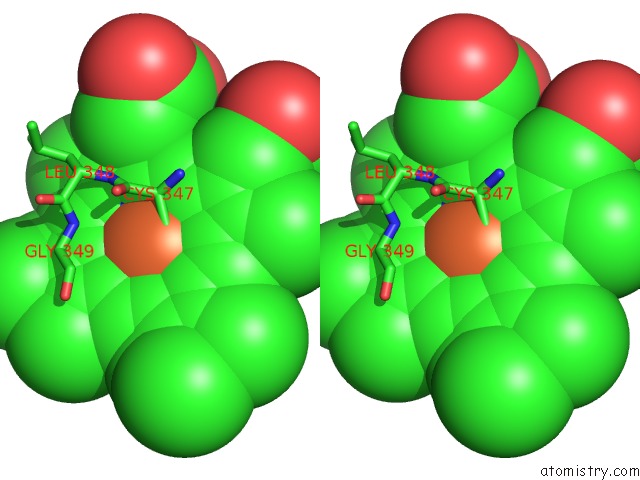
Stereo pair view
Mono view
Stereo pair view
A full contact list of Iron with other atoms in the Fe binding
site number 1 of Crystal Structure of Oxyb, A Cytochrome P450 Implicated in An Oxidative Phenol Coupling Reaction During Vancomycin Biosynthesis within 5.0Å range:
|
Reference:
K.Zerbe,
O.Pylypenko,
F.Vitali,
W.Zhang,
S.Rouset,
M.Heck,
J.W.Vrijbloed,
D.Bischoff,
B.Bister,
R.D.Sussmuth,
S.Pelzer,
W.Wohlleben,
J.A.Robinson,
I.Schlichting.
Crystal Structure of Oxyb, A Cytochrome P450 Implicated in An Oxidative Phenol Coupling Reaction During Vancomycin Biosynthesis. J.Biol.Chem. V. 277 47476 2002.
ISSN: ISSN 0021-9258
PubMed: 12207020
DOI: 10.1074/JBC.M206342200
Page generated: Sat Aug 3 09:45:53 2024
ISSN: ISSN 0021-9258
PubMed: 12207020
DOI: 10.1074/JBC.M206342200
Last articles
Zn in 9J0NZn in 9J0O
Zn in 9J0P
Zn in 9FJX
Zn in 9EKB
Zn in 9C0F
Zn in 9CAH
Zn in 9CH0
Zn in 9CH3
Zn in 9CH1