Iron »
PDB 1lfk-1lr6 »
1lkm »
Iron in PDB 1lkm: Crystal Structure of Desulfovibrio Vulgaris Rubrerythrin All-Iron(III) Form
Protein crystallography data
The structure of Crystal Structure of Desulfovibrio Vulgaris Rubrerythrin All-Iron(III) Form, PDB code: 1lkm
was solved by
S.Jin,
D.M.Kurtz Jr.,
Z.J.Liu,
J.Rose,
B.C.Wang,
with X-Ray Crystallography technique. A brief refinement statistics is given in the table below:
| Resolution Low / High (Å) | 25.95 / 1.69 |
| Space group | I 2 2 2 |
| Cell size a, b, c (Å), α, β, γ (°) | 48.877, 80.609, 100.084, 90.00, 90.00, 90.00 |
| R / Rfree (%) | 19 / 21 |
Iron Binding Sites:
The binding sites of Iron atom in the Crystal Structure of Desulfovibrio Vulgaris Rubrerythrin All-Iron(III) Form
(pdb code 1lkm). This binding sites where shown within
5.0 Angstroms radius around Iron atom.
In total 3 binding sites of Iron where determined in the Crystal Structure of Desulfovibrio Vulgaris Rubrerythrin All-Iron(III) Form, PDB code: 1lkm:
Jump to Iron binding site number: 1; 2; 3;
In total 3 binding sites of Iron where determined in the Crystal Structure of Desulfovibrio Vulgaris Rubrerythrin All-Iron(III) Form, PDB code: 1lkm:
Jump to Iron binding site number: 1; 2; 3;
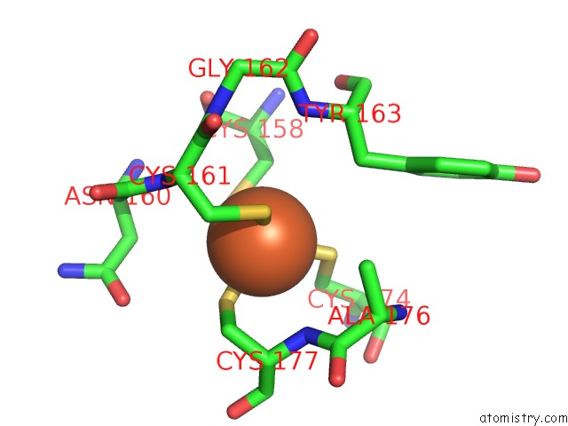
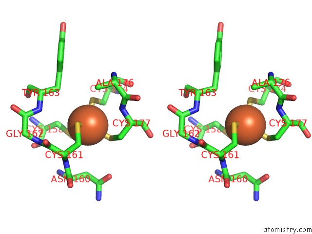
Iron binding site 1 out of 3 in 1lkm
Go back to
Iron binding site 1 out
of 3 in the Crystal Structure of Desulfovibrio Vulgaris Rubrerythrin All-Iron(III) Form
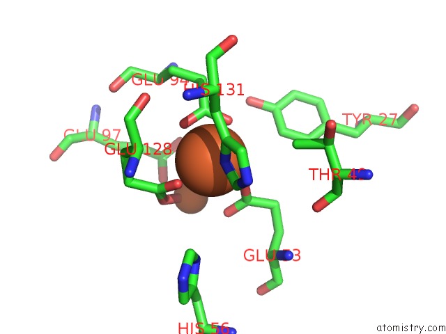
Mono view
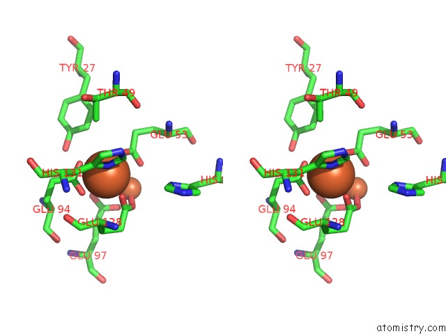
Stereo pair view
Mono view
Stereo pair view
A full contact list of Iron with other atoms in the Fe binding
site number 1 of Crystal Structure of Desulfovibrio Vulgaris Rubrerythrin All-Iron(III) Form within 5.0Å range:
|
Iron binding site 2 out of 3 in 1lkm
Go back to
Iron binding site 2 out
of 3 in the Crystal Structure of Desulfovibrio Vulgaris Rubrerythrin All-Iron(III) Form
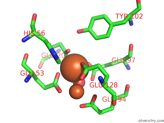
Mono view
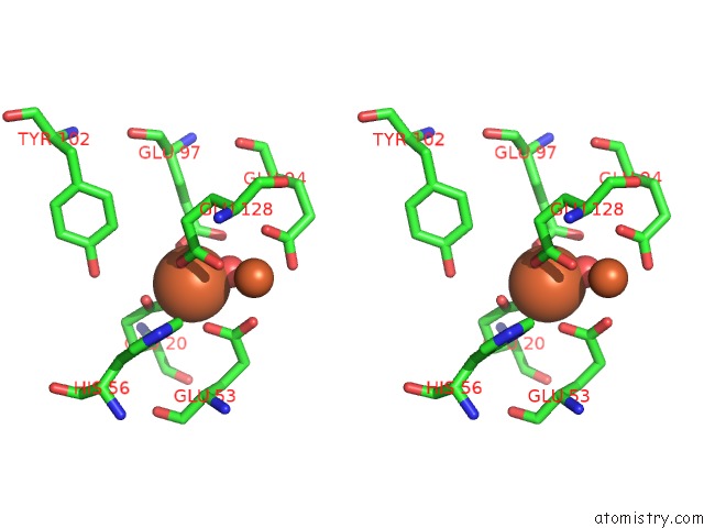
Stereo pair view
Mono view
Stereo pair view
A full contact list of Iron with other atoms in the Fe binding
site number 2 of Crystal Structure of Desulfovibrio Vulgaris Rubrerythrin All-Iron(III) Form within 5.0Å range:
|
Iron binding site 3 out of 3 in 1lkm
Go back to
Iron binding site 3 out
of 3 in the Crystal Structure of Desulfovibrio Vulgaris Rubrerythrin All-Iron(III) Form
Mono view
Stereo pair view
Mono view
Stereo pair view
A full contact list of Iron with other atoms in the Fe binding
site number 3 of Crystal Structure of Desulfovibrio Vulgaris Rubrerythrin All-Iron(III) Form within 5.0Å range:
|
Reference:
S.Jin,
D.M.Kurtz Jr.,
Z.J.Liu,
J.Rose,
B.C.Wang.
X-Ray Crystal Structures of Reduced Rubrerythrin and Its Azide Adduct: A Structure-Based Mechanism For A Non-Heme Diiron Peroxidase J.Am.Chem.Soc. V. 124 9845 2002.
ISSN: ISSN 0002-7863
PubMed: 12175244
DOI: 10.1021/JA026587U
Page generated: Sat Aug 3 09:48:56 2024
ISSN: ISSN 0002-7863
PubMed: 12175244
DOI: 10.1021/JA026587U
Last articles
Zn in 9J0NZn in 9J0O
Zn in 9J0P
Zn in 9FJX
Zn in 9EKB
Zn in 9C0F
Zn in 9CAH
Zn in 9CH0
Zn in 9CH3
Zn in 9CH1