Iron »
PDB 1lfk-1lr6 »
1llp »
Iron in PDB 1llp: Lignin Peroxidase (Isozyme H2) Pi 4.15
Protein crystallography data
The structure of Lignin Peroxidase (Isozyme H2) Pi 4.15, PDB code: 1llp
was solved by
T.H.Choinowski,
K.Piontek,
T.Glumoff,
with X-Ray Crystallography technique. A brief refinement statistics is given in the table below:
| Resolution Low / High (Å) | 10.00 / 1.70 |
| Space group | P 21 21 21 |
| Cell size a, b, c (Å), α, β, γ (°) | 60.700, 74.710, 106.350, 90.00, 90.00, 90.00 |
| R / Rfree (%) | n/a / n/a |
Other elements in 1llp:
The structure of Lignin Peroxidase (Isozyme H2) Pi 4.15 also contains other interesting chemical elements:
| Calcium | (Ca) | 2 atoms |
Iron Binding Sites:
The binding sites of Iron atom in the Lignin Peroxidase (Isozyme H2) Pi 4.15
(pdb code 1llp). This binding sites where shown within
5.0 Angstroms radius around Iron atom.
In total only one binding site of Iron was determined in the Lignin Peroxidase (Isozyme H2) Pi 4.15, PDB code: 1llp:
In total only one binding site of Iron was determined in the Lignin Peroxidase (Isozyme H2) Pi 4.15, PDB code: 1llp:
Iron binding site 1 out of 1 in 1llp
Go back to
Iron binding site 1 out
of 1 in the Lignin Peroxidase (Isozyme H2) Pi 4.15
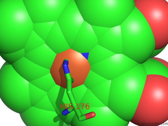
Mono view
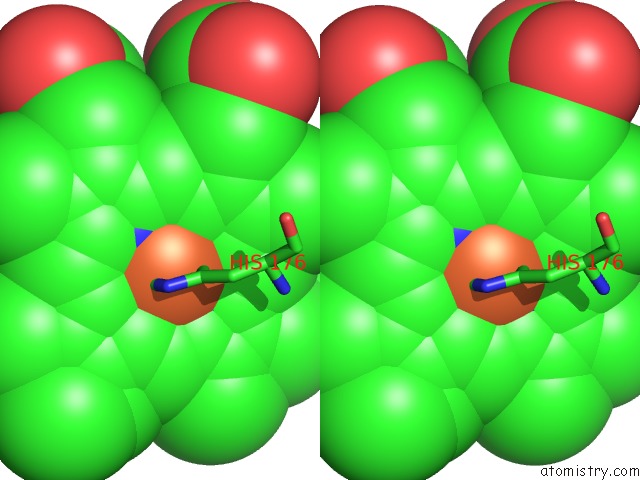
Stereo pair view
Mono view
Stereo pair view
A full contact list of Iron with other atoms in the Fe binding
site number 1 of Lignin Peroxidase (Isozyme H2) Pi 4.15 within 5.0Å range:
|
Reference:
T.Choinowski,
W.Blodig,
K.H.Winterhalter,
K.Piontek.
The Crystal Structure of Lignin Peroxidase at 1.70 A Resolution Reveals A Hydroxy Group on the Cbeta of Tryptophan 171: A Novel Radical Site Formed During the Redox Cycle. J.Mol.Biol. V. 286 809 1999.
ISSN: ISSN 0022-2836
PubMed: 10024453
DOI: 10.1006/JMBI.1998.2507
Page generated: Wed Jul 16 17:27:01 2025
ISSN: ISSN 0022-2836
PubMed: 10024453
DOI: 10.1006/JMBI.1998.2507
Last articles
Fe in 2YXOFe in 2YRS
Fe in 2YXC
Fe in 2YNM
Fe in 2YVJ
Fe in 2YP1
Fe in 2YU2
Fe in 2YU1
Fe in 2YQB
Fe in 2YOO