Iron »
PDB 1lfk-1lr6 »
1lm4 »
Iron in PDB 1lm4: Structure of Peptide Deformylase From Staphylococcus Aureus at 1.45 A
Enzymatic activity of Structure of Peptide Deformylase From Staphylococcus Aureus at 1.45 A
All present enzymatic activity of Structure of Peptide Deformylase From Staphylococcus Aureus at 1.45 A:
3.5.1.88;
3.5.1.88;
Protein crystallography data
The structure of Structure of Peptide Deformylase From Staphylococcus Aureus at 1.45 A, PDB code: 1lm4
was solved by
A.Kreusch,
G.Spraggon,
C.C.Lee,
H.Klock,
D.Mcmullan,
K.Ng,
T.Shin,
J.Vincent,
I.Warner,
C.Ericson,
S.A.Lesley,
with X-Ray Crystallography technique. A brief refinement statistics is given in the table below:
| Resolution Low / High (Å) | 45.26 / 1.45 |
| Space group | P 21 21 21 |
| Cell size a, b, c (Å), α, β, γ (°) | 38.224, 76.703, 112.116, 90.00, 90.00, 90.00 |
| R / Rfree (%) | 17 / 20 |
Iron Binding Sites:
The binding sites of Iron atom in the Structure of Peptide Deformylase From Staphylococcus Aureus at 1.45 A
(pdb code 1lm4). This binding sites where shown within
5.0 Angstroms radius around Iron atom.
In total 2 binding sites of Iron where determined in the Structure of Peptide Deformylase From Staphylococcus Aureus at 1.45 A, PDB code: 1lm4:
Jump to Iron binding site number: 1; 2;
In total 2 binding sites of Iron where determined in the Structure of Peptide Deformylase From Staphylococcus Aureus at 1.45 A, PDB code: 1lm4:
Jump to Iron binding site number: 1; 2;
Iron binding site 1 out of 2 in 1lm4
Go back to
Iron binding site 1 out
of 2 in the Structure of Peptide Deformylase From Staphylococcus Aureus at 1.45 A
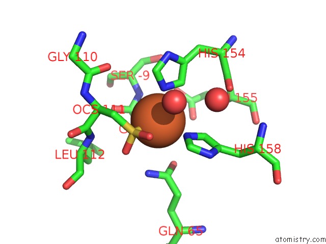
Mono view

Stereo pair view
Mono view
Stereo pair view
A full contact list of Iron with other atoms in the Fe binding
site number 1 of Structure of Peptide Deformylase From Staphylococcus Aureus at 1.45 A within 5.0Å range:
|
Iron binding site 2 out of 2 in 1lm4
Go back to
Iron binding site 2 out
of 2 in the Structure of Peptide Deformylase From Staphylococcus Aureus at 1.45 A
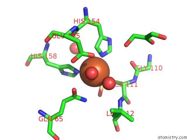
Mono view
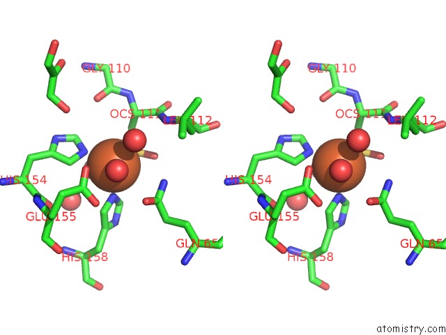
Stereo pair view
Mono view
Stereo pair view
A full contact list of Iron with other atoms in the Fe binding
site number 2 of Structure of Peptide Deformylase From Staphylococcus Aureus at 1.45 A within 5.0Å range:
|
Reference:
A.Kreusch,
G.Spraggon,
C.C.Lee,
H.Klock,
D.Mcmullan,
K.Ng,
T.Shin,
J.Vincent,
I.Warner,
C.Ericson,
S.A.Lesley.
Structure Analysis of Peptide Deformylases From Streptococcus Pneumoniae,Staphylococcus Aureus, Thermotoga Maritima, and Pseudomonas Aeruginosa: Snapshots of the Oxygen Sensitivity of Peptide Deformylase J.Mol.Biol. V. 330 309 2003.
ISSN: ISSN 0022-2836
PubMed: 12823970
DOI: 10.1016/S0022-2836(03)00596-5
Page generated: Sat Aug 3 09:52:08 2024
ISSN: ISSN 0022-2836
PubMed: 12823970
DOI: 10.1016/S0022-2836(03)00596-5
Last articles
Zn in 9J0NZn in 9J0O
Zn in 9J0P
Zn in 9FJX
Zn in 9EKB
Zn in 9C0F
Zn in 9CAH
Zn in 9CH0
Zn in 9CH3
Zn in 9CH1