Iron »
PDB 1lfk-1lr6 »
1lr6 »
Iron in PDB 1lr6: Crystal Structure of V45Y Mutant of Cytochrome B5
Protein crystallography data
The structure of Crystal Structure of V45Y Mutant of Cytochrome B5, PDB code: 1lr6
was solved by
J.-H.Gan,
J.Wu,
Z.-Q.Wang,
Y.-H.Wang,
Z.-X.Huang,
Z.-X.Xia,
with X-Ray Crystallography technique. A brief refinement statistics is given in the table below:
| Resolution Low / High (Å) | 22.82 / 1.90 |
| Space group | C 1 2 1 |
| Cell size a, b, c (Å), α, β, γ (°) | 69.404, 40.441, 39.196, 90.00, 111.70, 90.00 |
| R / Rfree (%) | 19.1 / 23.6 |
Iron Binding Sites:
The binding sites of Iron atom in the Crystal Structure of V45Y Mutant of Cytochrome B5
(pdb code 1lr6). This binding sites where shown within
5.0 Angstroms radius around Iron atom.
In total only one binding site of Iron was determined in the Crystal Structure of V45Y Mutant of Cytochrome B5, PDB code: 1lr6:
In total only one binding site of Iron was determined in the Crystal Structure of V45Y Mutant of Cytochrome B5, PDB code: 1lr6:
Iron binding site 1 out of 1 in 1lr6
Go back to
Iron binding site 1 out
of 1 in the Crystal Structure of V45Y Mutant of Cytochrome B5
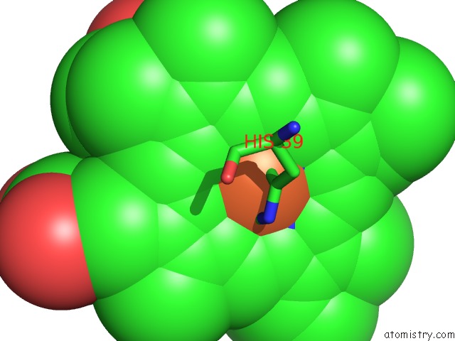
Mono view
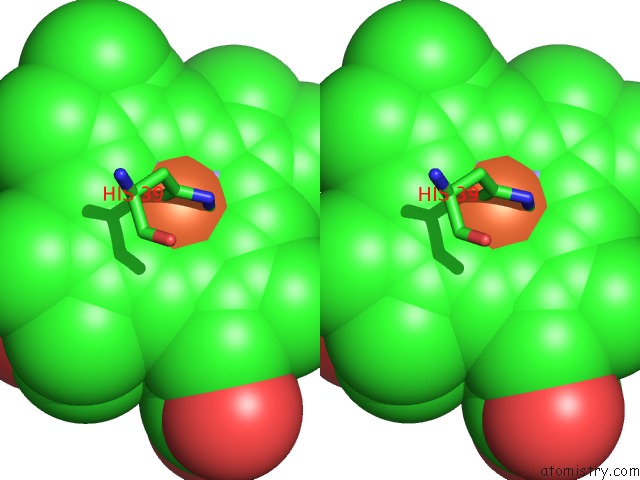
Stereo pair view
Mono view
Stereo pair view
A full contact list of Iron with other atoms in the Fe binding
site number 1 of Crystal Structure of V45Y Mutant of Cytochrome B5 within 5.0Å range:
|
Reference:
J.H.Gan,
J.Wu,
Z.Q.Wang,
Y.H.Wang,
Z.X.Huang,
Z.X.Xia.
Structures of V45E and V45Y Mutants and Structure Comparison of A Variety of Cytochrome B5 Mutants. Acta Crystallogr.,Sect.D V. 58 1298 2002.
ISSN: ISSN 0907-4449
PubMed: 12136141
DOI: 10.1107/S0907444902010016
Page generated: Sat Aug 3 09:53:59 2024
ISSN: ISSN 0907-4449
PubMed: 12136141
DOI: 10.1107/S0907444902010016
Last articles
Zn in 9J0NZn in 9J0O
Zn in 9J0P
Zn in 9FJX
Zn in 9EKB
Zn in 9C0F
Zn in 9CAH
Zn in 9CH0
Zn in 9CH3
Zn in 9CH1