Iron »
PDB 1lrm-1m6m »
1m56 »
Iron in PDB 1m56: Structure of Cytochrome C Oxidase From Rhodobactor Sphaeroides (Wild Type)
Enzymatic activity of Structure of Cytochrome C Oxidase From Rhodobactor Sphaeroides (Wild Type)
All present enzymatic activity of Structure of Cytochrome C Oxidase From Rhodobactor Sphaeroides (Wild Type):
1.9.3.1;
1.9.3.1;
Protein crystallography data
The structure of Structure of Cytochrome C Oxidase From Rhodobactor Sphaeroides (Wild Type), PDB code: 1m56
was solved by
M.Svensson-Ek,
J.Abramson,
G.Larsson,
S.Tornroth,
P.Brezezinski,
S.Iwata,
with X-Ray Crystallography technique. A brief refinement statistics is given in the table below:
| Resolution Low / High (Å) | 40.00 / 2.30 |
| Space group | H 3 |
| Cell size a, b, c (Å), α, β, γ (°) | 340.360, 340.360, 89.668, 90.00, 90.00, 120.00 |
| R / Rfree (%) | 23.6 / 27.5 |
Other elements in 1m56:
The structure of Structure of Cytochrome C Oxidase From Rhodobactor Sphaeroides (Wild Type) also contains other interesting chemical elements:
| Magnesium | (Mg) | 2 atoms |
| Copper | (Cu) | 6 atoms |
| Calcium | (Ca) | 2 atoms |
Iron Binding Sites:
The binding sites of Iron atom in the Structure of Cytochrome C Oxidase From Rhodobactor Sphaeroides (Wild Type)
(pdb code 1m56). This binding sites where shown within
5.0 Angstroms radius around Iron atom.
In total 4 binding sites of Iron where determined in the Structure of Cytochrome C Oxidase From Rhodobactor Sphaeroides (Wild Type), PDB code: 1m56:
Jump to Iron binding site number: 1; 2; 3; 4;
In total 4 binding sites of Iron where determined in the Structure of Cytochrome C Oxidase From Rhodobactor Sphaeroides (Wild Type), PDB code: 1m56:
Jump to Iron binding site number: 1; 2; 3; 4;
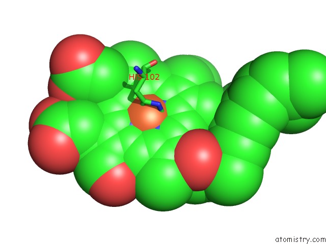
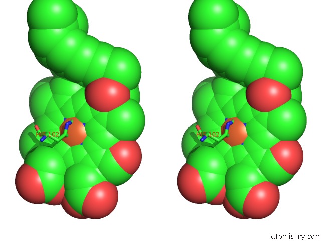
Iron binding site 1 out of 4 in 1m56
Go back to
Iron binding site 1 out
of 4 in the Structure of Cytochrome C Oxidase From Rhodobactor Sphaeroides (Wild Type)
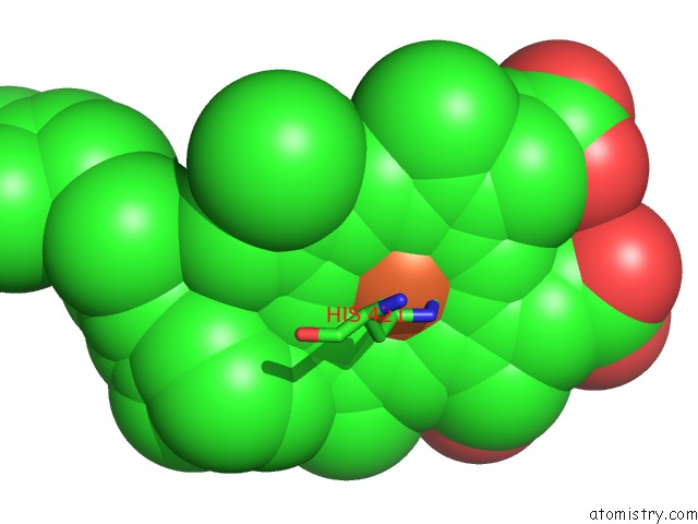
Mono view
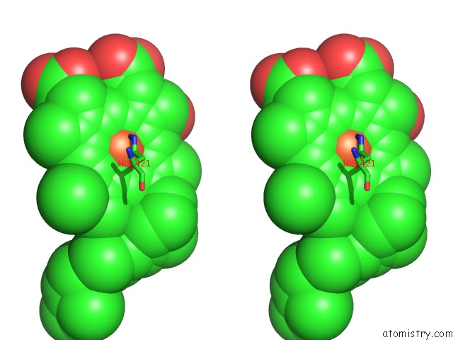
Stereo pair view
Mono view
Stereo pair view
A full contact list of Iron with other atoms in the Fe binding
site number 1 of Structure of Cytochrome C Oxidase From Rhodobactor Sphaeroides (Wild Type) within 5.0Å range:
|
Iron binding site 2 out of 4 in 1m56
Go back to
Iron binding site 2 out
of 4 in the Structure of Cytochrome C Oxidase From Rhodobactor Sphaeroides (Wild Type)
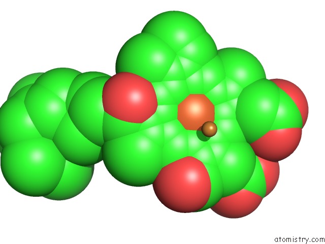
Mono view
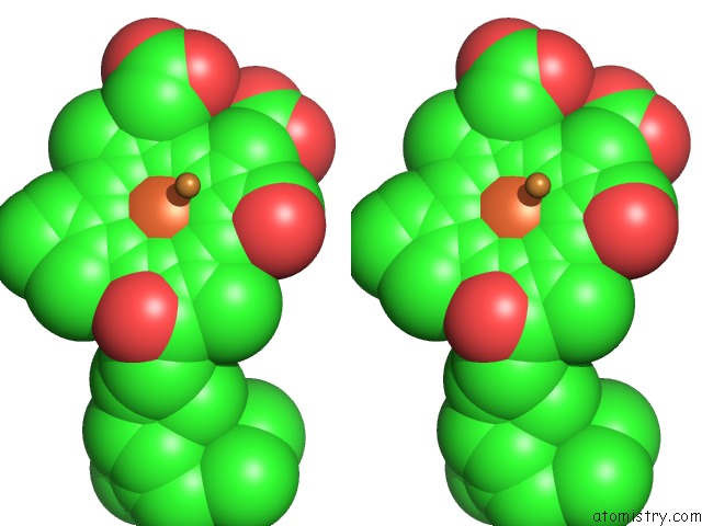
Stereo pair view
Mono view
Stereo pair view
A full contact list of Iron with other atoms in the Fe binding
site number 2 of Structure of Cytochrome C Oxidase From Rhodobactor Sphaeroides (Wild Type) within 5.0Å range:
|
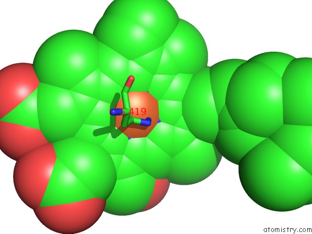
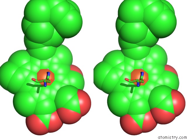
Iron binding site 3 out of 4 in 1m56
Go back to
Iron binding site 3 out
of 4 in the Structure of Cytochrome C Oxidase From Rhodobactor Sphaeroides (Wild Type)
Mono view
Stereo pair view
Mono view
Stereo pair view
A full contact list of Iron with other atoms in the Fe binding
site number 3 of Structure of Cytochrome C Oxidase From Rhodobactor Sphaeroides (Wild Type) within 5.0Å range:
|
Iron binding site 4 out of 4 in 1m56
Go back to
Iron binding site 4 out
of 4 in the Structure of Cytochrome C Oxidase From Rhodobactor Sphaeroides (Wild Type)
Mono view
Stereo pair view
Mono view
Stereo pair view
A full contact list of Iron with other atoms in the Fe binding
site number 4 of Structure of Cytochrome C Oxidase From Rhodobactor Sphaeroides (Wild Type) within 5.0Å range:
|
Reference:
M.Svensson-Ek,
J.Abramson,
G.Larsson,
S.Tornroth,
P.Brzezinski,
S.Iwata.
The X-Ray Crystal Structures of Wild-Type and Eq(I-286) Mutant Cytochrome C Oxidases From Rhodobacter Sphaeroides. J.Mol.Biol. V. 321 329 2002.
ISSN: ISSN 0022-2836
PubMed: 12144789
DOI: 10.1016/S0022-2836(02)00619-8
Page generated: Wed Jul 16 17:40:31 2025
ISSN: ISSN 0022-2836
PubMed: 12144789
DOI: 10.1016/S0022-2836(02)00619-8
Last articles
Fe in 2YXOFe in 2YRS
Fe in 2YXC
Fe in 2YNM
Fe in 2YVJ
Fe in 2YP1
Fe in 2YU2
Fe in 2YU1
Fe in 2YQB
Fe in 2YOO