Iron »
PDB 1m6z-1mko »
1mhz »
Iron in PDB 1mhz: Methane Monooxygenase Hydroxylase
Enzymatic activity of Methane Monooxygenase Hydroxylase
All present enzymatic activity of Methane Monooxygenase Hydroxylase:
1.14.13.25;
1.14.13.25;
Protein crystallography data
The structure of Methane Monooxygenase Hydroxylase, PDB code: 1mhz
was solved by
N.Elango,
R.Radhakrishnan,
W.A.Froland,
B.J.Waller,
C.A.Earhart,
J.D.Lipscomb,
D.H.Ohlendorf,
with X-Ray Crystallography technique. A brief refinement statistics is given in the table below:
| Resolution Low / High (Å) | 5.00 / 2.70 |
| Space group | C 2 2 21 |
| Cell size a, b, c (Å), α, β, γ (°) | 293.380, 64.010, 143.650, 90.00, 90.00, 90.00 |
| R / Rfree (%) | 15.2 / n/a |
Iron Binding Sites:
The binding sites of Iron atom in the Methane Monooxygenase Hydroxylase
(pdb code 1mhz). This binding sites where shown within
5.0 Angstroms radius around Iron atom.
In total 2 binding sites of Iron where determined in the Methane Monooxygenase Hydroxylase, PDB code: 1mhz:
Jump to Iron binding site number: 1; 2;
In total 2 binding sites of Iron where determined in the Methane Monooxygenase Hydroxylase, PDB code: 1mhz:
Jump to Iron binding site number: 1; 2;
Iron binding site 1 out of 2 in 1mhz
Go back to
Iron binding site 1 out
of 2 in the Methane Monooxygenase Hydroxylase

Mono view
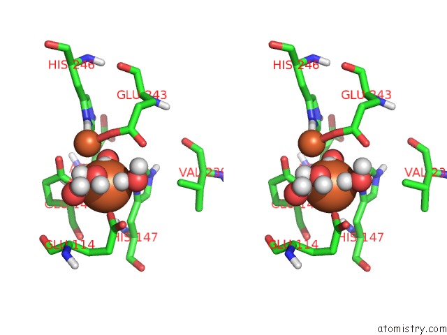
Stereo pair view
Mono view
Stereo pair view
A full contact list of Iron with other atoms in the Fe binding
site number 1 of Methane Monooxygenase Hydroxylase within 5.0Å range:
|
Iron binding site 2 out of 2 in 1mhz
Go back to
Iron binding site 2 out
of 2 in the Methane Monooxygenase Hydroxylase
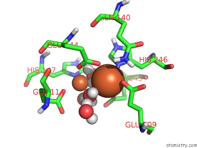
Mono view

Stereo pair view
Mono view
Stereo pair view
A full contact list of Iron with other atoms in the Fe binding
site number 2 of Methane Monooxygenase Hydroxylase within 5.0Å range:
|
Reference:
N.Elango,
R.Radhakrishnan,
W.A.Froland,
B.J.Wallar,
C.A.Earhart,
J.D.Lipscomb,
D.H.Ohlendorf.
Crystal Structure of the Hydroxylase Component of Methane Monooxygenase From Methylosinus Trichosporium OB3B Protein Sci. V. 6 556 1997.
ISSN: ISSN 0961-8368
PubMed: 9070438
Page generated: Wed Jul 16 18:03:40 2025
ISSN: ISSN 0961-8368
PubMed: 9070438
Last articles
Fe in 2FWLFe in 2FW5
Fe in 2FRZ
Fe in 2FRK
Fe in 2FRJ
Fe in 2FRI
Fe in 2FRF
Fe in 2FRC
Fe in 2FR7
Fe in 2FLA