Iron »
PDB 1mkq-1mpw »
1mmt »
Iron in PDB 1mmt: Crystal Structure of Ternary Complex of the Catalytic Domain of Human Phenylalanine Hydroxylase (Fe(II)) Complexed with Tetrahydrobiopterin and Norleucine
Enzymatic activity of Crystal Structure of Ternary Complex of the Catalytic Domain of Human Phenylalanine Hydroxylase (Fe(II)) Complexed with Tetrahydrobiopterin and Norleucine
All present enzymatic activity of Crystal Structure of Ternary Complex of the Catalytic Domain of Human Phenylalanine Hydroxylase (Fe(II)) Complexed with Tetrahydrobiopterin and Norleucine:
1.14.16.1;
1.14.16.1;
Protein crystallography data
The structure of Crystal Structure of Ternary Complex of the Catalytic Domain of Human Phenylalanine Hydroxylase (Fe(II)) Complexed with Tetrahydrobiopterin and Norleucine, PDB code: 1mmt
was solved by
O.A.Andersen,
T.Flatmark,
E.Hough,
with X-Ray Crystallography technique. A brief refinement statistics is given in the table below:
| Resolution Low / High (Å) | 10.00 / 2.00 |
| Space group | C 2 2 21 |
| Cell size a, b, c (Å), α, β, γ (°) | 65.140, 106.638, 123.611, 90.00, 90.00, 90.00 |
| R / Rfree (%) | 21.3 / 24.3 |
Iron Binding Sites:
The binding sites of Iron atom in the Crystal Structure of Ternary Complex of the Catalytic Domain of Human Phenylalanine Hydroxylase (Fe(II)) Complexed with Tetrahydrobiopterin and Norleucine
(pdb code 1mmt). This binding sites where shown within
5.0 Angstroms radius around Iron atom.
In total only one binding site of Iron was determined in the Crystal Structure of Ternary Complex of the Catalytic Domain of Human Phenylalanine Hydroxylase (Fe(II)) Complexed with Tetrahydrobiopterin and Norleucine, PDB code: 1mmt:
In total only one binding site of Iron was determined in the Crystal Structure of Ternary Complex of the Catalytic Domain of Human Phenylalanine Hydroxylase (Fe(II)) Complexed with Tetrahydrobiopterin and Norleucine, PDB code: 1mmt:
Iron binding site 1 out of 1 in 1mmt
Go back to
Iron binding site 1 out
of 1 in the Crystal Structure of Ternary Complex of the Catalytic Domain of Human Phenylalanine Hydroxylase (Fe(II)) Complexed with Tetrahydrobiopterin and Norleucine
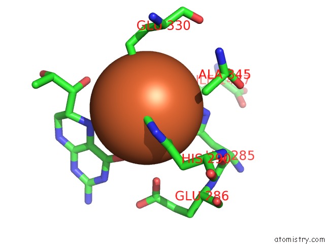
Mono view
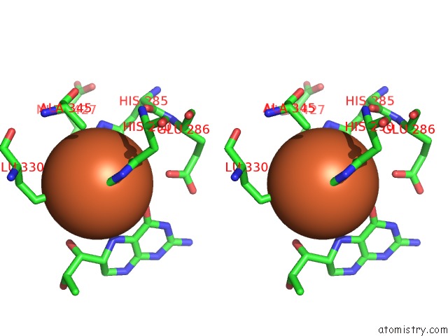
Stereo pair view
Mono view
Stereo pair view
A full contact list of Iron with other atoms in the Fe binding
site number 1 of Crystal Structure of Ternary Complex of the Catalytic Domain of Human Phenylalanine Hydroxylase (Fe(II)) Complexed with Tetrahydrobiopterin and Norleucine within 5.0Å range:
|
Reference:
O.A.Andersen,
A.J.Stokka,
T.Flatmark,
E.Hough.
2.0A Resolution Crystal Structures of the Ternary Complexes of Human Phenylalanine Hydroxylase Catalytic Domain with Tetrahydrobiopterin and 3-(2-Thienyl)-L-Alanine or L-Norleucine: Substrate Specificity and Molecular Motions Related to Substrate Binding J.Mol.Biol. V. 333 747 2003.
ISSN: ISSN 0022-2836
PubMed: 14568534
DOI: 10.1016/J.JMB.2003.09.004
Page generated: Sat Aug 3 11:02:14 2024
ISSN: ISSN 0022-2836
PubMed: 14568534
DOI: 10.1016/J.JMB.2003.09.004
Last articles
Zn in 9J0NZn in 9J0O
Zn in 9J0P
Zn in 9FJX
Zn in 9EKB
Zn in 9C0F
Zn in 9CAH
Zn in 9CH0
Zn in 9CH3
Zn in 9CH1