Iron »
PDB 1mpy-1n5u »
1mty »
Iron in PDB 1mty: Methane Monooxygenase Hydroxylase From Methylococcus Capsulatus (Bath)
Enzymatic activity of Methane Monooxygenase Hydroxylase From Methylococcus Capsulatus (Bath)
All present enzymatic activity of Methane Monooxygenase Hydroxylase From Methylococcus Capsulatus (Bath):
1.14.13.25;
1.14.13.25;
Protein crystallography data
The structure of Methane Monooxygenase Hydroxylase From Methylococcus Capsulatus (Bath), PDB code: 1mty
was solved by
A.C.Rosenzweig,
P.Nordlund,
S.J.Lippard,
C.A.Frederick,
with X-Ray Crystallography technique. A brief refinement statistics is given in the table below:
| Resolution Low / High (Å) | 5.00 / 1.70 |
| Space group | P 21 21 21 |
| Cell size a, b, c (Å), α, β, γ (°) | 61.700, 109.600, 330.200, 90.00, 90.00, 90.00 |
| R / Rfree (%) | 18.3 / n/a |
Iron Binding Sites:
The binding sites of Iron atom in the Methane Monooxygenase Hydroxylase From Methylococcus Capsulatus (Bath)
(pdb code 1mty). This binding sites where shown within
5.0 Angstroms radius around Iron atom.
In total 4 binding sites of Iron where determined in the Methane Monooxygenase Hydroxylase From Methylococcus Capsulatus (Bath), PDB code: 1mty:
Jump to Iron binding site number: 1; 2; 3; 4;
In total 4 binding sites of Iron where determined in the Methane Monooxygenase Hydroxylase From Methylococcus Capsulatus (Bath), PDB code: 1mty:
Jump to Iron binding site number: 1; 2; 3; 4;
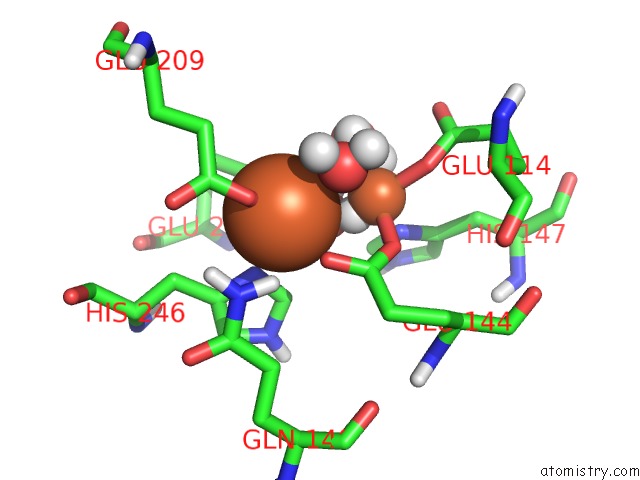
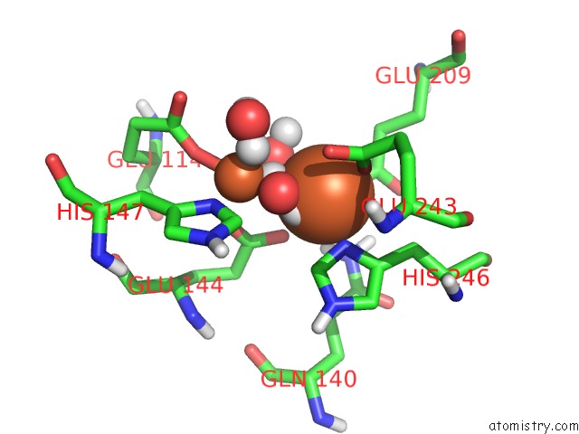
Iron binding site 1 out of 4 in 1mty
Go back to
Iron binding site 1 out
of 4 in the Methane Monooxygenase Hydroxylase From Methylococcus Capsulatus (Bath)
Mono view
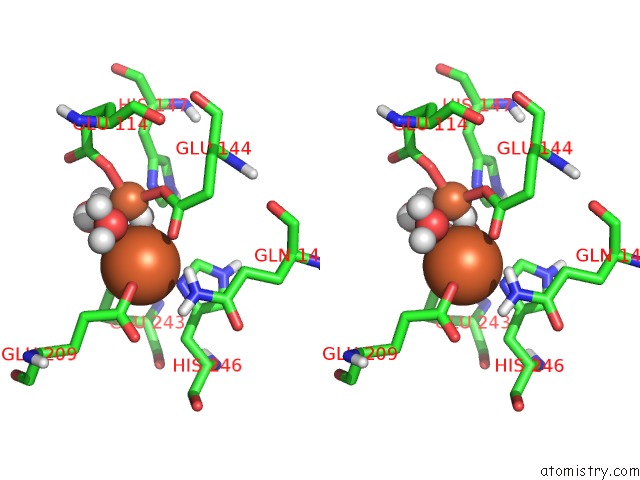
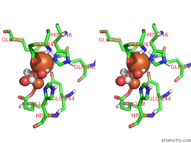
Stereo pair view
Mono view
Stereo pair view
A full contact list of Iron with other atoms in the Fe binding
site number 1 of Methane Monooxygenase Hydroxylase From Methylococcus Capsulatus (Bath) within 5.0Å range:
|
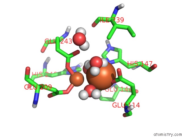
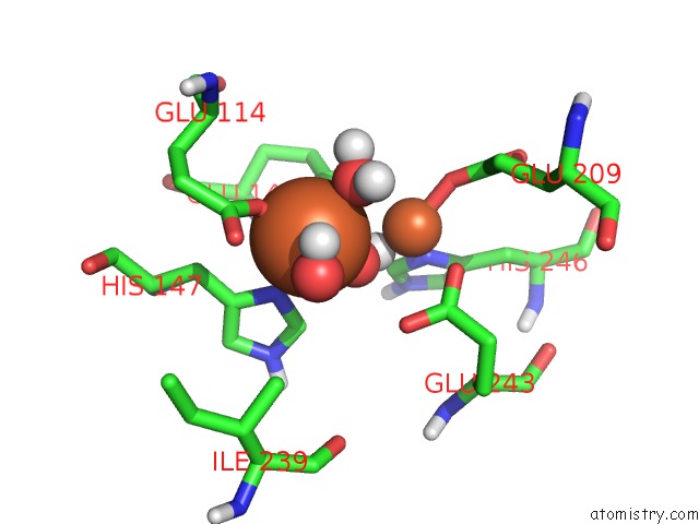
Iron binding site 2 out of 4 in 1mty
Go back to
Iron binding site 2 out
of 4 in the Methane Monooxygenase Hydroxylase From Methylococcus Capsulatus (Bath)
Mono view
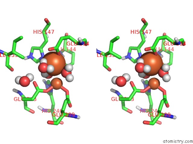
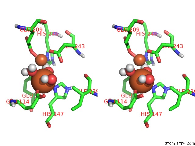
Stereo pair view
Mono view
Stereo pair view
A full contact list of Iron with other atoms in the Fe binding
site number 2 of Methane Monooxygenase Hydroxylase From Methylococcus Capsulatus (Bath) within 5.0Å range:
|
Iron binding site 3 out of 4 in 1mty
Go back to
Iron binding site 3 out
of 4 in the Methane Monooxygenase Hydroxylase From Methylococcus Capsulatus (Bath)
Mono view
Stereo pair view
Mono view
Stereo pair view
A full contact list of Iron with other atoms in the Fe binding
site number 3 of Methane Monooxygenase Hydroxylase From Methylococcus Capsulatus (Bath) within 5.0Å range:
|
Iron binding site 4 out of 4 in 1mty
Go back to
Iron binding site 4 out
of 4 in the Methane Monooxygenase Hydroxylase From Methylococcus Capsulatus (Bath)
Mono view
Stereo pair view
Mono view
Stereo pair view
A full contact list of Iron with other atoms in the Fe binding
site number 4 of Methane Monooxygenase Hydroxylase From Methylococcus Capsulatus (Bath) within 5.0Å range:
|
Reference:
A.C.Rosenzweig,
H.Brandstetter,
D.A.Whittington,
P.Nordlund,
S.J.Lippard,
C.A.Frederick.
Crystal Structures of the Methane Monooxygenase Hydroxylase From Methylococcus Capsulatus (Bath): Implications For Substrate Gating and Component Interactions. Proteins V. 29 141 1997.
ISSN: ISSN 0887-3585
PubMed: 9329079
DOI: 10.1002/(SICI)1097-0134(199710)29:2<141::AID-PROT2>3.3.CO;2-W
Page generated: Wed Jul 16 18:18:22 2025
ISSN: ISSN 0887-3585
PubMed: 9329079
DOI: 10.1002/(SICI)1097-0134(199710)29:2<141::AID-PROT2>3.3.CO;2-W
Last articles
Fe in 1YQPFe in 1YTC
Fe in 1YRC
Fe in 1YRD
Fe in 1YQ9
Fe in 1YQ4
Fe in 1YQ3
Fe in 1YQO
Fe in 1YHU
Fe in 1YNR