Iron »
PDB 1mpy-1n5u »
1mwb »
Iron in PDB 1mwb: Solution Structure of the Recombinant Hemoglobin From the Cyanobacterium Synechocystis Sp. Pcc 6803 in Its Hemichrome State
Iron Binding Sites:
The binding sites of Iron atom in the Solution Structure of the Recombinant Hemoglobin From the Cyanobacterium Synechocystis Sp. Pcc 6803 in Its Hemichrome State
(pdb code 1mwb). This binding sites where shown within
5.0 Angstroms radius around Iron atom.
In total only one binding site of Iron was determined in the Solution Structure of the Recombinant Hemoglobin From the Cyanobacterium Synechocystis Sp. Pcc 6803 in Its Hemichrome State, PDB code: 1mwb:
In total only one binding site of Iron was determined in the Solution Structure of the Recombinant Hemoglobin From the Cyanobacterium Synechocystis Sp. Pcc 6803 in Its Hemichrome State, PDB code: 1mwb:
Iron binding site 1 out of 1 in 1mwb
Go back to
Iron binding site 1 out
of 1 in the Solution Structure of the Recombinant Hemoglobin From the Cyanobacterium Synechocystis Sp. Pcc 6803 in Its Hemichrome State
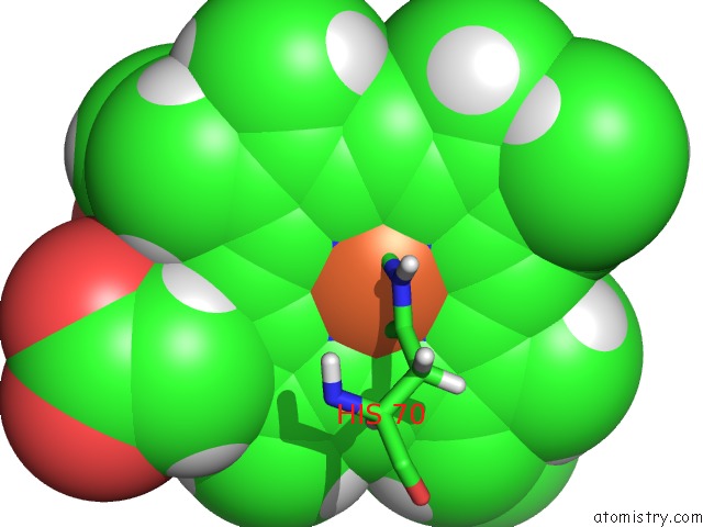
Mono view
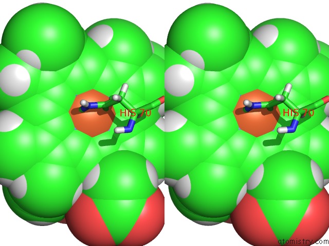
Stereo pair view
Mono view
Stereo pair view
A full contact list of Iron with other atoms in the Fe binding
site number 1 of Solution Structure of the Recombinant Hemoglobin From the Cyanobacterium Synechocystis Sp. Pcc 6803 in Its Hemichrome State within 5.0Å range:
|
Reference:
C.J.Falzone,
B.C.Vu,
N.L.Scott,
J.T.Lecomte.
The Solution Structure of the Recombinant Hemoglobin From the Cyanobacterium Synechocystis Sp. Pcc 6803 in Its Hemichrome State J.Mol.Biol. V. 324 1015 2002.
ISSN: ISSN 0022-2836
PubMed: 12470956
DOI: 10.1016/S0022-2836(02)01093-8
Page generated: Wed Jul 16 18:19:42 2025
ISSN: ISSN 0022-2836
PubMed: 12470956
DOI: 10.1016/S0022-2836(02)01093-8
Last articles
Fe in 2EXVFe in 2EUU
Fe in 2EUT
Fe in 2EVP
Fe in 2EVK
Fe in 2EUS
Fe in 2EUR
Fe in 2E84
Fe in 2EUQ
Fe in 2ET0