Iron »
PDB 1mpy-1n5u »
1mwd »
Iron in PDB 1mwd: Wild Type Deoxy Myoglobin
Protein crystallography data
The structure of Wild Type Deoxy Myoglobin, PDB code: 1mwd
was solved by
G.N.Murshudov,
S.Krzywda,
A.M.Brzozowski,
M.Jaskolski,
E.E.Scott,
A.A.Klizas,
Q.H.Gibson,
J.S.Olson,
A.J.Wilkinson,
with X-Ray Crystallography technique. A brief refinement statistics is given in the table below:
| Resolution Low / High (Å) | 24.00 / 1.80 |
| Space group | I 1 21 1 |
| Cell size a, b, c (Å), α, β, γ (°) | 122.120, 42.140, 91.490, 90.00, 92.85, 90.00 |
| R / Rfree (%) | 19.6 / 24.3 |
Iron Binding Sites:
The binding sites of Iron atom in the Wild Type Deoxy Myoglobin
(pdb code 1mwd). This binding sites where shown within
5.0 Angstroms radius around Iron atom.
In total 2 binding sites of Iron where determined in the Wild Type Deoxy Myoglobin, PDB code: 1mwd:
Jump to Iron binding site number: 1; 2;
In total 2 binding sites of Iron where determined in the Wild Type Deoxy Myoglobin, PDB code: 1mwd:
Jump to Iron binding site number: 1; 2;
Iron binding site 1 out of 2 in 1mwd
Go back to
Iron binding site 1 out
of 2 in the Wild Type Deoxy Myoglobin
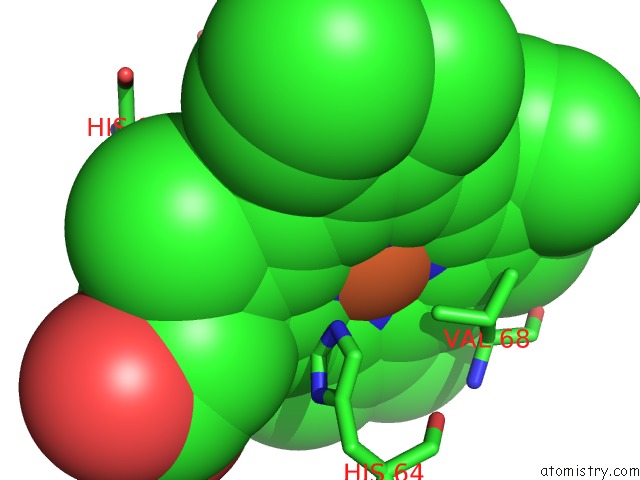
Mono view
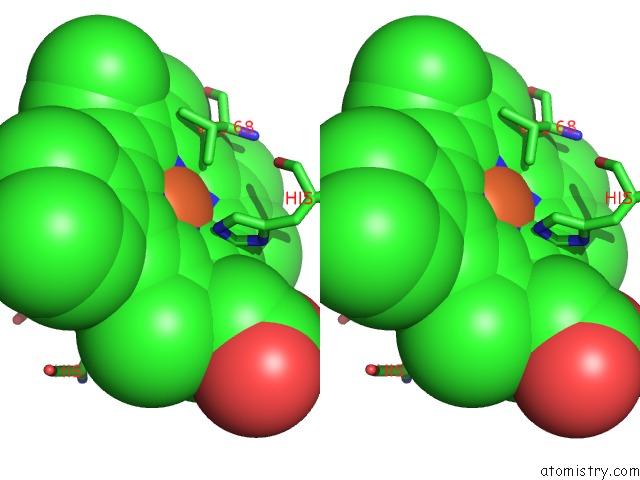
Stereo pair view
Mono view
Stereo pair view
A full contact list of Iron with other atoms in the Fe binding
site number 1 of Wild Type Deoxy Myoglobin within 5.0Å range:
|
Iron binding site 2 out of 2 in 1mwd
Go back to
Iron binding site 2 out
of 2 in the Wild Type Deoxy Myoglobin
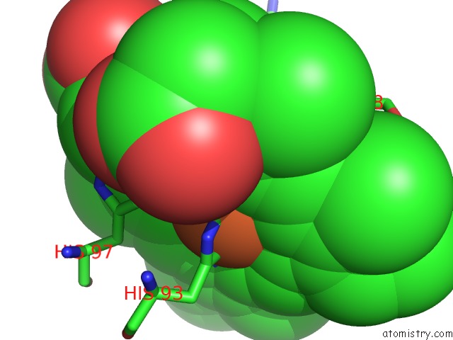
Mono view
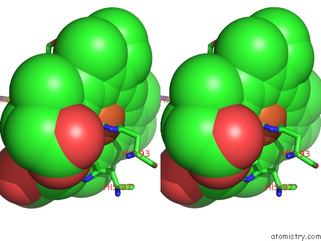
Stereo pair view
Mono view
Stereo pair view
A full contact list of Iron with other atoms in the Fe binding
site number 2 of Wild Type Deoxy Myoglobin within 5.0Å range:
|
Reference:
S.Krzywda,
G.N.Murshudov,
A.M.Brzozowski,
M.Jaskolski,
E.E.Scott,
S.A.Klizas,
Q.H.Gibson,
J.S.Olson,
A.J.Wilkinson.
Stabilizing Bound O2 in Myoglobin By VALINE68 (E11) to Asparagine Substitution. Biochemistry V. 37 15896 1998.
ISSN: ISSN 0006-2960
PubMed: 9843395
DOI: 10.1021/BI9812470
Page generated: Sat Aug 3 11:08:49 2024
ISSN: ISSN 0006-2960
PubMed: 9843395
DOI: 10.1021/BI9812470
Last articles
Cl in 8C14Cl in 8C1I
Cl in 8C1G
Cl in 8C1F
Cl in 8C1E
Cl in 8C1D
Cl in 8C15
Cl in 8C04
Cl in 8C0K
Cl in 8BZW