Iron »
PDB 1mpy-1n5u »
1myj »
Iron in PDB 1myj: Distal Polarity in Ligand Binding to Myoglobin: Structural and Functional Characterization of A THREONINE68(E11) Mutant
Protein crystallography data
The structure of Distal Polarity in Ligand Binding to Myoglobin: Structural and Functional Characterization of A THREONINE68(E11) Mutant, PDB code: 1myj
was solved by
S.J.Smerdon,
T.J.Oldfield,
A.J.Wilkinson,
Z.Dauter,
K.Petratos,
K.S.Wilson,
with X-Ray Crystallography technique. A brief refinement statistics is given in the table below:
| Resolution Low / High (Å) | 7.00 / 1.90 |
| Space group | I 21 |
| Cell size a, b, c (Å), α, β, γ (°) | 123.750, 42.480, 92.050, 90.00, 92.05, 90.00 |
| R / Rfree (%) | n/a / n/a |
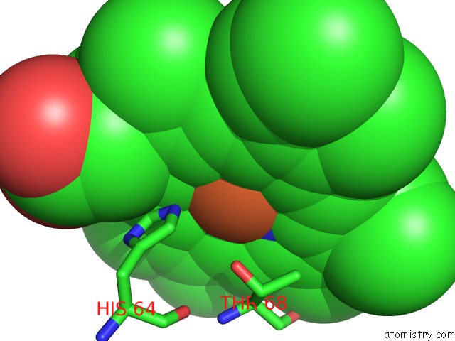
Iron Binding Sites:
The binding sites of Iron atom in the Distal Polarity in Ligand Binding to Myoglobin: Structural and Functional Characterization of A THREONINE68(E11) Mutant
(pdb code 1myj). This binding sites where shown within
5.0 Angstroms radius around Iron atom.
In total 2 binding sites of Iron where determined in the Distal Polarity in Ligand Binding to Myoglobin: Structural and Functional Characterization of A THREONINE68(E11) Mutant, PDB code: 1myj:
Jump to Iron binding site number: 1; 2;
In total 2 binding sites of Iron where determined in the Distal Polarity in Ligand Binding to Myoglobin: Structural and Functional Characterization of A THREONINE68(E11) Mutant, PDB code: 1myj:
Jump to Iron binding site number: 1; 2;
Iron binding site 1 out of 2 in 1myj
Go back to
Iron binding site 1 out
of 2 in the Distal Polarity in Ligand Binding to Myoglobin: Structural and Functional Characterization of A THREONINE68(E11) Mutant
Mono view

Stereo pair view
Mono view
Stereo pair view
A full contact list of Iron with other atoms in the Fe binding
site number 1 of Distal Polarity in Ligand Binding to Myoglobin: Structural and Functional Characterization of A THREONINE68(E11) Mutant within 5.0Å range:
|
Iron binding site 2 out of 2 in 1myj
Go back to
Iron binding site 2 out
of 2 in the Distal Polarity in Ligand Binding to Myoglobin: Structural and Functional Characterization of A THREONINE68(E11) Mutant

Mono view

Stereo pair view
Mono view
Stereo pair view
A full contact list of Iron with other atoms in the Fe binding
site number 2 of Distal Polarity in Ligand Binding to Myoglobin: Structural and Functional Characterization of A THREONINE68(E11) Mutant within 5.0Å range:
|
Reference:
S.J.Smerdon,
G.G.Dodson,
A.J.Wilkinson,
Q.H.Gibson,
R.S.Blackmore,
T.E.Carver,
J.S.Olson.
Distal Pocket Polarity in Ligand Binding to Myoglobin: Structural and Functional Characterization of A THREONINE68(E11) Mutant. Biochemistry V. 30 6252 1991.
ISSN: ISSN 0006-2960
PubMed: 1905570
DOI: 10.1021/BI00239A025
Page generated: Sat Aug 3 11:11:21 2024
ISSN: ISSN 0006-2960
PubMed: 1905570
DOI: 10.1021/BI00239A025
Last articles
Zn in 9MJ5Zn in 9HNW
Zn in 9G0L
Zn in 9FNE
Zn in 9DZN
Zn in 9E0I
Zn in 9D32
Zn in 9DAK
Zn in 8ZXC
Zn in 8ZUF