Iron »
PDB 1mpy-1n5u »
1myt »
Iron in PDB 1myt: Crystal Structure to 1.74 Angstroms Resolution of Metmyoglobin From Yellowfin Tuna (Thunnus Albacares): An Example of A Myoglobin Lacking the D Helix
Protein crystallography data
The structure of Crystal Structure to 1.74 Angstroms Resolution of Metmyoglobin From Yellowfin Tuna (Thunnus Albacares): An Example of A Myoglobin Lacking the D Helix, PDB code: 1myt
was solved by
G.I.Birnbaum,
S.V.Evans,
M.Przybylska,
D.R.Rose,
with X-Ray Crystallography technique. A brief refinement statistics is given in the table below:
| Resolution Low / High (Å) | 6.00 / 1.74 |
| Space group | P 21 21 21 |
| Cell size a, b, c (Å), α, β, γ (°) | 44.497, 72.318, 52.221, 90.00, 90.00, 90.00 |
| R / Rfree (%) | n/a / n/a |
Iron Binding Sites:
The binding sites of Iron atom in the Crystal Structure to 1.74 Angstroms Resolution of Metmyoglobin From Yellowfin Tuna (Thunnus Albacares): An Example of A Myoglobin Lacking the D Helix
(pdb code 1myt). This binding sites where shown within
5.0 Angstroms radius around Iron atom.
In total only one binding site of Iron was determined in the Crystal Structure to 1.74 Angstroms Resolution of Metmyoglobin From Yellowfin Tuna (Thunnus Albacares): An Example of A Myoglobin Lacking the D Helix, PDB code: 1myt:
In total only one binding site of Iron was determined in the Crystal Structure to 1.74 Angstroms Resolution of Metmyoglobin From Yellowfin Tuna (Thunnus Albacares): An Example of A Myoglobin Lacking the D Helix, PDB code: 1myt:
Iron binding site 1 out of 1 in 1myt
Go back to
Iron binding site 1 out
of 1 in the Crystal Structure to 1.74 Angstroms Resolution of Metmyoglobin From Yellowfin Tuna (Thunnus Albacares): An Example of A Myoglobin Lacking the D Helix
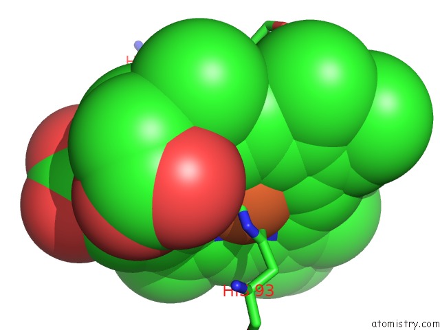
Mono view
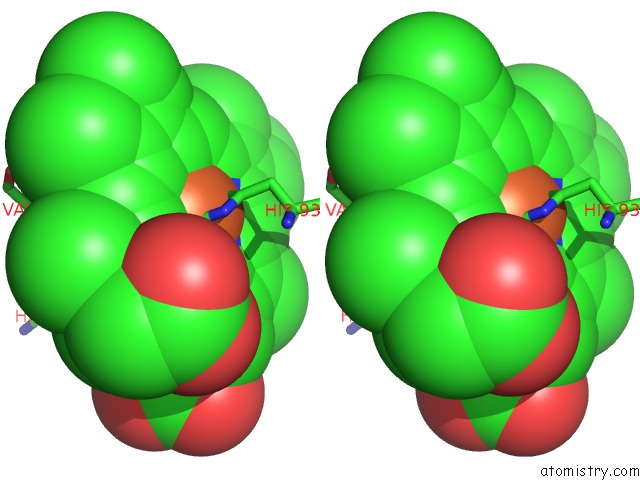
Stereo pair view
Mono view
Stereo pair view
A full contact list of Iron with other atoms in the Fe binding
site number 1 of Crystal Structure to 1.74 Angstroms Resolution of Metmyoglobin From Yellowfin Tuna (Thunnus Albacares): An Example of A Myoglobin Lacking the D Helix within 5.0Å range:
|
Reference:
G.I.Birnbaum,
S.V.Evans,
M.Przybylska,
D.R.Rose.
1.70 A Resolution Structure of Myoglobin From Yellowfin Tuna. An Example of A Myoglobin Lacking the D Helix. Acta Crystallogr.,Sect.D V. 50 283 1994.
ISSN: ISSN 0907-4449
PubMed: 15299440
DOI: 10.1107/S0907444993014271
Page generated: Wed Jul 16 18:21:26 2025
ISSN: ISSN 0907-4449
PubMed: 15299440
DOI: 10.1107/S0907444993014271
Last articles
Fe in 2YXOFe in 2YRS
Fe in 2YXC
Fe in 2YNM
Fe in 2YVJ
Fe in 2YP1
Fe in 2YU2
Fe in 2YU1
Fe in 2YQB
Fe in 2YOO