Iron »
PDB 1mpy-1n5u »
1myz »
Iron in PDB 1myz: Co Complex of Myoglobin Mb-Yqr at Rt Solved From Laue Data.
Protein crystallography data
The structure of Co Complex of Myoglobin Mb-Yqr at Rt Solved From Laue Data., PDB code: 1myz
was solved by
D.Bourgeois,
B.Vallone,
F.Schotte,
A.Arcovito,
A.E.Miele,
G.Sciara,
M.Wulff,
P.Anfinrud,
M.Brunori,
with X-Ray Crystallography technique. A brief refinement statistics is given in the table below:
| Resolution Low / High (Å) | 29.88 / 1.60 |
| Space group | P 6 |
| Cell size a, b, c (Å), α, β, γ (°) | 91.200, 91.200, 45.712, 90.00, 90.00, 120.00 |
| R / Rfree (%) | 16.6 / 18.4 |
Iron Binding Sites:
The binding sites of Iron atom in the Co Complex of Myoglobin Mb-Yqr at Rt Solved From Laue Data.
(pdb code 1myz). This binding sites where shown within
5.0 Angstroms radius around Iron atom.
In total only one binding site of Iron was determined in the Co Complex of Myoglobin Mb-Yqr at Rt Solved From Laue Data., PDB code: 1myz:
In total only one binding site of Iron was determined in the Co Complex of Myoglobin Mb-Yqr at Rt Solved From Laue Data., PDB code: 1myz:
Iron binding site 1 out of 1 in 1myz
Go back to
Iron binding site 1 out
of 1 in the Co Complex of Myoglobin Mb-Yqr at Rt Solved From Laue Data.
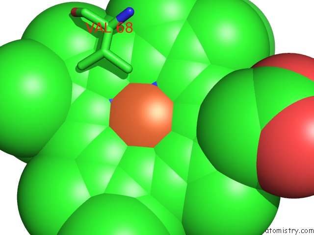
Mono view
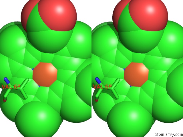
Stereo pair view
Mono view
Stereo pair view
A full contact list of Iron with other atoms in the Fe binding
site number 1 of Co Complex of Myoglobin Mb-Yqr at Rt Solved From Laue Data. within 5.0Å range:
|
Reference:
D.Bourgeois,
B.Vallone,
F.Schotte,
A.Arcovito,
A.E.Miele,
G.Sciara,
M.Wulff,
P.Anfinrud,
M.Brunori.
Complex Landscape of Protein Structural Dynamics Unveiled By Nanosecond Laue Crystallography. Proc.Natl.Acad.Sci.Usa V. 100 8704 2003.
ISSN: ISSN 0027-8424
PubMed: 12847289
DOI: 10.1073/PNAS.1430900100
Page generated: Wed Jul 16 18:21:36 2025
ISSN: ISSN 0027-8424
PubMed: 12847289
DOI: 10.1073/PNAS.1430900100
Last articles
Fe in 2YXOFe in 2YRS
Fe in 2YXC
Fe in 2YNM
Fe in 2YVJ
Fe in 2YP1
Fe in 2YU2
Fe in 2YU1
Fe in 2YQB
Fe in 2YOO