Iron »
PDB 1n5w-1nmi »
1n5w »
Iron in PDB 1n5w: Crystal Structure of the Cu,Mo-Co Dehydrogenase (Codh); Oxidized Form
Enzymatic activity of Crystal Structure of the Cu,Mo-Co Dehydrogenase (Codh); Oxidized Form
All present enzymatic activity of Crystal Structure of the Cu,Mo-Co Dehydrogenase (Codh); Oxidized Form:
1.2.99.2;
1.2.99.2;
Protein crystallography data
The structure of Crystal Structure of the Cu,Mo-Co Dehydrogenase (Codh); Oxidized Form, PDB code: 1n5w
was solved by
H.Dobbek,
L.Gremer,
R.Kiefersauer,
R.Huber,
O.Meyer,
with X-Ray Crystallography technique. A brief refinement statistics is given in the table below:
| Resolution Low / High (Å) | 18.00 / 1.50 |
| Space group | P 21 21 21 |
| Cell size a, b, c (Å), α, β, γ (°) | 119.295, 132.088, 159.825, 90.00, 90.00, 90.00 |
| R / Rfree (%) | 13.5 / 17.1 |
Other elements in 1n5w:
The structure of Crystal Structure of the Cu,Mo-Co Dehydrogenase (Codh); Oxidized Form also contains other interesting chemical elements:
| Molybdenum | (Mo) | 2 atoms |
| Copper | (Cu) | 2 atoms |
Iron Binding Sites:
The binding sites of Iron atom in the Crystal Structure of the Cu,Mo-Co Dehydrogenase (Codh); Oxidized Form
(pdb code 1n5w). This binding sites where shown within
5.0 Angstroms radius around Iron atom.
In total 8 binding sites of Iron where determined in the Crystal Structure of the Cu,Mo-Co Dehydrogenase (Codh); Oxidized Form, PDB code: 1n5w:
Jump to Iron binding site number: 1; 2; 3; 4; 5; 6; 7; 8;
In total 8 binding sites of Iron where determined in the Crystal Structure of the Cu,Mo-Co Dehydrogenase (Codh); Oxidized Form, PDB code: 1n5w:
Jump to Iron binding site number: 1; 2; 3; 4; 5; 6; 7; 8;
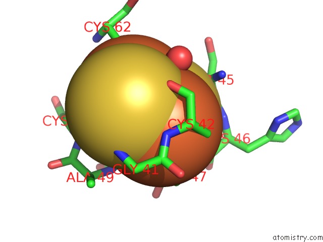
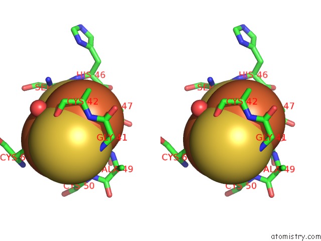
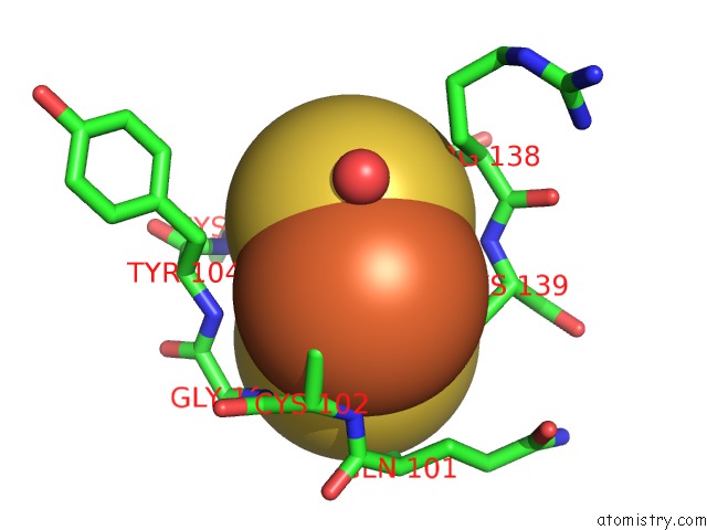
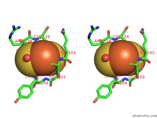
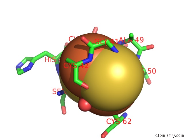
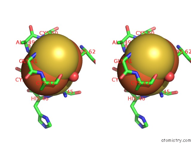
Iron binding site 1 out of 8 in 1n5w
Go back to
Iron binding site 1 out
of 8 in the Crystal Structure of the Cu,Mo-Co Dehydrogenase (Codh); Oxidized Form
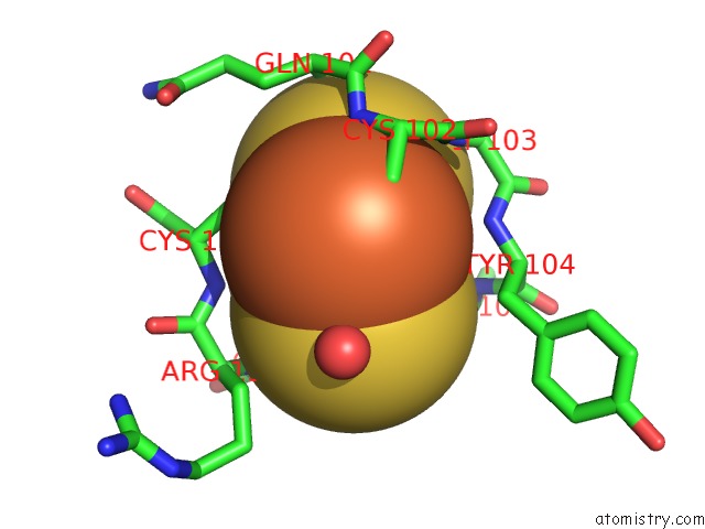
Mono view
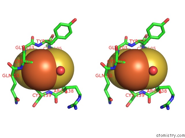
Stereo pair view
Mono view
Stereo pair view
A full contact list of Iron with other atoms in the Fe binding
site number 1 of Crystal Structure of the Cu,Mo-Co Dehydrogenase (Codh); Oxidized Form within 5.0Å range:
|
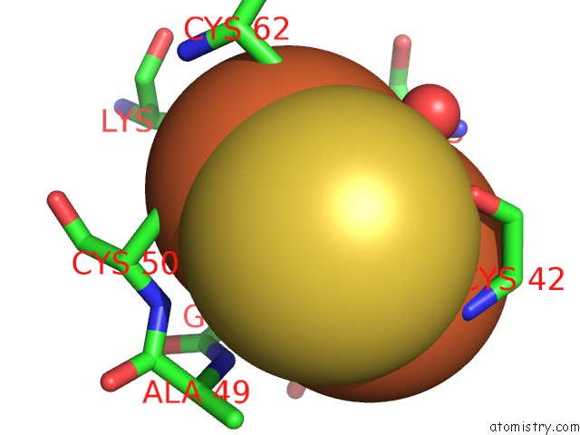
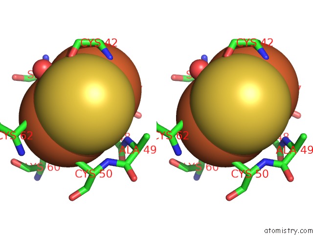
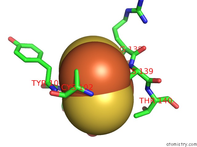
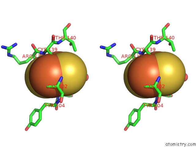
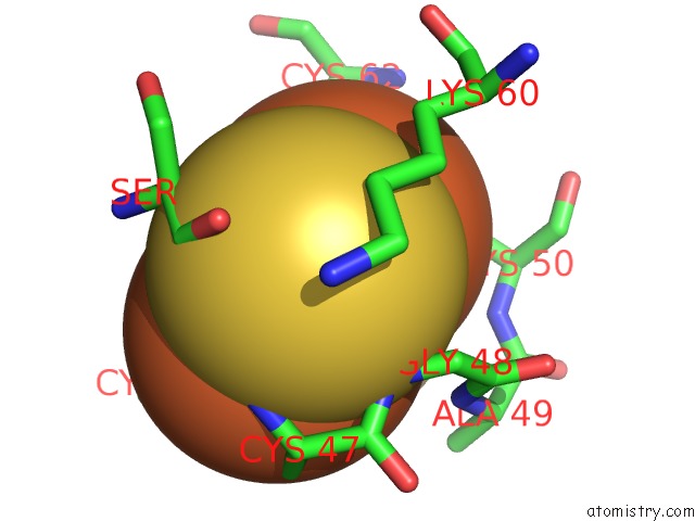
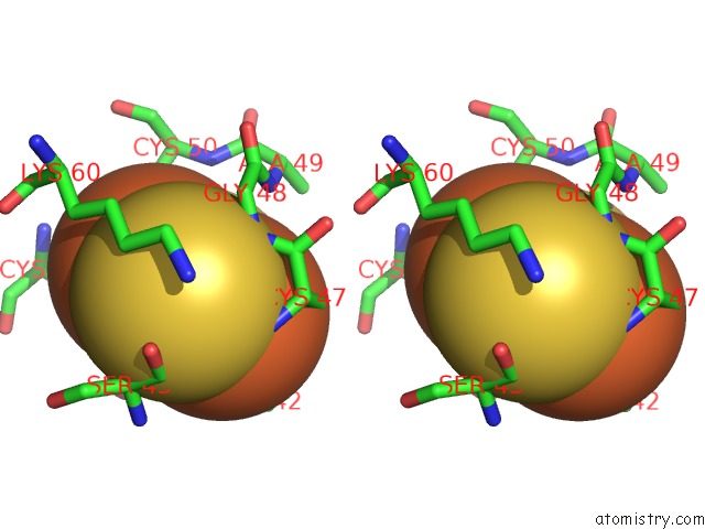
Iron binding site 2 out of 8 in 1n5w
Go back to
Iron binding site 2 out
of 8 in the Crystal Structure of the Cu,Mo-Co Dehydrogenase (Codh); Oxidized Form
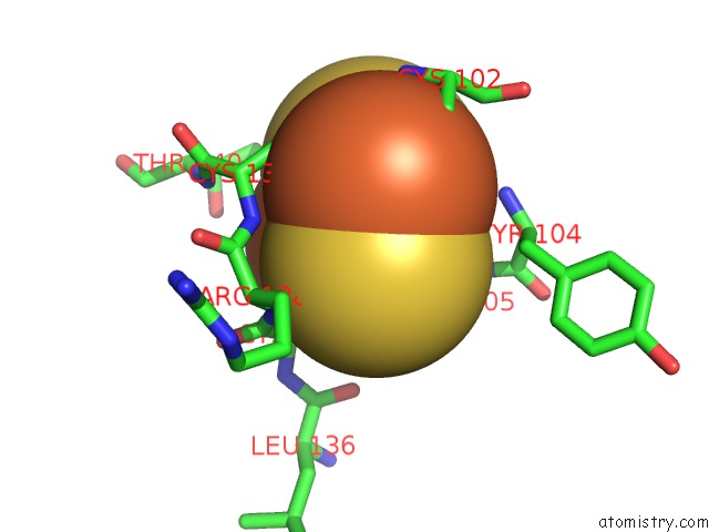
Mono view
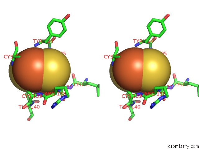
Stereo pair view
Mono view
Stereo pair view
A full contact list of Iron with other atoms in the Fe binding
site number 2 of Crystal Structure of the Cu,Mo-Co Dehydrogenase (Codh); Oxidized Form within 5.0Å range:
|
Iron binding site 3 out of 8 in 1n5w
Go back to
Iron binding site 3 out
of 8 in the Crystal Structure of the Cu,Mo-Co Dehydrogenase (Codh); Oxidized Form
Mono view
Stereo pair view
Mono view
Stereo pair view
A full contact list of Iron with other atoms in the Fe binding
site number 3 of Crystal Structure of the Cu,Mo-Co Dehydrogenase (Codh); Oxidized Form within 5.0Å range:
|
Iron binding site 4 out of 8 in 1n5w
Go back to
Iron binding site 4 out
of 8 in the Crystal Structure of the Cu,Mo-Co Dehydrogenase (Codh); Oxidized Form
Mono view
Stereo pair view
Mono view
Stereo pair view
A full contact list of Iron with other atoms in the Fe binding
site number 4 of Crystal Structure of the Cu,Mo-Co Dehydrogenase (Codh); Oxidized Form within 5.0Å range:
|
Iron binding site 5 out of 8 in 1n5w
Go back to
Iron binding site 5 out
of 8 in the Crystal Structure of the Cu,Mo-Co Dehydrogenase (Codh); Oxidized Form
Mono view
Stereo pair view
Mono view
Stereo pair view
A full contact list of Iron with other atoms in the Fe binding
site number 5 of Crystal Structure of the Cu,Mo-Co Dehydrogenase (Codh); Oxidized Form within 5.0Å range:
|
Iron binding site 6 out of 8 in 1n5w
Go back to
Iron binding site 6 out
of 8 in the Crystal Structure of the Cu,Mo-Co Dehydrogenase (Codh); Oxidized Form
Mono view
Stereo pair view
Mono view
Stereo pair view
A full contact list of Iron with other atoms in the Fe binding
site number 6 of Crystal Structure of the Cu,Mo-Co Dehydrogenase (Codh); Oxidized Form within 5.0Å range:
|
Iron binding site 7 out of 8 in 1n5w
Go back to
Iron binding site 7 out
of 8 in the Crystal Structure of the Cu,Mo-Co Dehydrogenase (Codh); Oxidized Form
Mono view
Stereo pair view
Mono view
Stereo pair view
A full contact list of Iron with other atoms in the Fe binding
site number 7 of Crystal Structure of the Cu,Mo-Co Dehydrogenase (Codh); Oxidized Form within 5.0Å range:
|
Iron binding site 8 out of 8 in 1n5w
Go back to
Iron binding site 8 out
of 8 in the Crystal Structure of the Cu,Mo-Co Dehydrogenase (Codh); Oxidized Form
Mono view
Stereo pair view
Mono view
Stereo pair view
A full contact list of Iron with other atoms in the Fe binding
site number 8 of Crystal Structure of the Cu,Mo-Co Dehydrogenase (Codh); Oxidized Form within 5.0Å range:
|
Reference:
H.Dobbek,
L.Gremer,
R.Kiefersauer,
R.Huber,
O.Meyer.
Catalysis at A Dinuclear [Cusmo(=O)Oh] Cluster in A Co Dehydrogenase Resolved at 1.1-A Resolution Proc.Natl.Acad.Sci.Usa V. 99 15971 2002.
ISSN: ISSN 0027-8424
PubMed: 12475995
DOI: 10.1073/PNAS.212640899
Page generated: Sat Aug 3 11:25:13 2024
ISSN: ISSN 0027-8424
PubMed: 12475995
DOI: 10.1073/PNAS.212640899
Last articles
Zn in 9J0NZn in 9J0O
Zn in 9J0P
Zn in 9FJX
Zn in 9EKB
Zn in 9C0F
Zn in 9CAH
Zn in 9CH0
Zn in 9CH3
Zn in 9CH1