Iron in PDB 1n60: Crystal Structure of the Cu,Mo-Co Dehydrogenase (Codh); Cyanide- Inactivated Form
Enzymatic activity of Crystal Structure of the Cu,Mo-Co Dehydrogenase (Codh); Cyanide- Inactivated Form
All present enzymatic activity of Crystal Structure of the Cu,Mo-Co Dehydrogenase (Codh); Cyanide- Inactivated Form:
1.2.99.2;
Protein crystallography data
The structure of Crystal Structure of the Cu,Mo-Co Dehydrogenase (Codh); Cyanide- Inactivated Form, PDB code: 1n60
was solved by
H.Dobbek,
L.Gremer,
R.Kiefersauer,
R.Huber,
O.Meyer,
with X-Ray Crystallography technique. A brief refinement statistics is given in the table below:
Resolution Low / High (Å)
|
17.80 /
1.19
|
Space group
|
P 21 21 21
|
Cell size a, b, c (Å), α, β, γ (°)
|
118.566,
130.642,
158.492,
90.00,
90.00,
90.00
|
R / Rfree (%)
|
14.2 /
17.1
|
Other elements in 1n60:
The structure of Crystal Structure of the Cu,Mo-Co Dehydrogenase (Codh); Cyanide- Inactivated Form also contains other interesting chemical elements:
Iron Binding Sites:
The binding sites of Iron atom in the Crystal Structure of the Cu,Mo-Co Dehydrogenase (Codh); Cyanide- Inactivated Form
(pdb code 1n60). This binding sites where shown within
5.0 Angstroms radius around Iron atom.
In total 8 binding sites of Iron where determined in the
Crystal Structure of the Cu,Mo-Co Dehydrogenase (Codh); Cyanide- Inactivated Form, PDB code: 1n60:
Jump to Iron binding site number:
1;
2;
3;
4;
5;
6;
7;
8;
Iron binding site 1 out
of 8 in 1n60
Go back to
Iron Binding Sites List in 1n60
Iron binding site 1 out
of 8 in the Crystal Structure of the Cu,Mo-Co Dehydrogenase (Codh); Cyanide- Inactivated Form
 Mono view
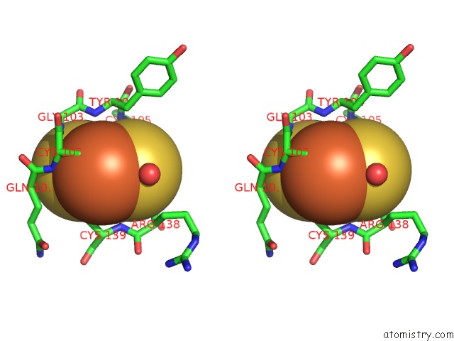
 Stereo pair view
|
A full contact list of Iron with other atoms in the Fe binding
site number 1 of Crystal Structure of the Cu,Mo-Co Dehydrogenase (Codh); Cyanide- Inactivated Form within 5.0Å range:
probe
|
atom
|
residue
|
distance (Å)
|
B
|
Occ
|
A:Fe4907
b:10.0
occ:1.00
|
FE1
|
A:FES4907
|
0.0
|
10.0
|
1.0
|
S2
|
A:FES4907
|
2.2
|
9.7
|
1.0
|
S1
|
A:FES4907
|
2.3
|
9.7
|
1.0
|
SG
|
A:CYS102
|
2.3
|
9.2
|
1.0
|
SG
|
A:CYS139
|
2.3
|
9.7
|
1.0
|
FE2
|
A:FES4907
|
2.7
|
9.9
|
1.0
|
CB
|
A:CYS139
|
3.3
|
10.7
|
1.0
|
CB
|
A:CYS102
|
3.4
|
10.2
|
1.0
|
N
|
A:CYS102
|
3.5
|
9.4
|
1.0
|
CA
|
A:CYS102
|
3.9
|
9.4
|
1.0
|
O
|
B:HOH4934
|
3.9
|
11.7
|
1.0
|
N
|
A:GLY103
|
3.9
|
9.4
|
1.0
|
N
|
A:CYS139
|
4.0
|
9.9
|
1.0
|
N
|
A:TYR104
|
4.2
|
9.9
|
1.0
|
C
|
A:CYS102
|
4.3
|
10.1
|
1.0
|
CA
|
A:CYS139
|
4.3
|
10.4
|
1.0
|
SG
|
A:CYS137
|
4.3
|
9.3
|
1.0
|
C
|
A:GLN101
|
4.6
|
9.5
|
1.0
|
SG
|
A:CYS105
|
4.7
|
9.9
|
1.0
|
N
|
A:CYS105
|
4.7
|
9.5
|
1.0
|
N
|
A:ARG138
|
4.8
|
9.6
|
1.0
|
N
|
A:GLN101
|
4.8
|
9.7
|
1.0
|
CB
|
A:TYR104
|
4.8
|
10.4
|
1.0
|
CA
|
A:GLY103
|
4.9
|
9.9
|
1.0
|
CB
|
A:GLN101
|
4.9
|
10.1
|
1.0
|
C
|
A:ARG138
|
4.9
|
9.9
|
1.0
|
C
|
A:GLY103
|
5.0
|
10.4
|
1.0
|
CA
|
A:TYR104
|
5.0
|
10.5
|
1.0
|
|
Iron binding site 2 out
of 8 in 1n60
Go back to
Iron Binding Sites List in 1n60
Iron binding site 2 out
of 8 in the Crystal Structure of the Cu,Mo-Co Dehydrogenase (Codh); Cyanide- Inactivated Form
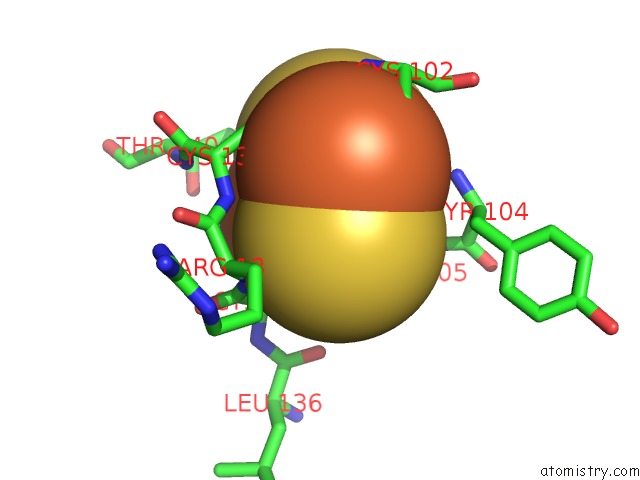
 Mono view
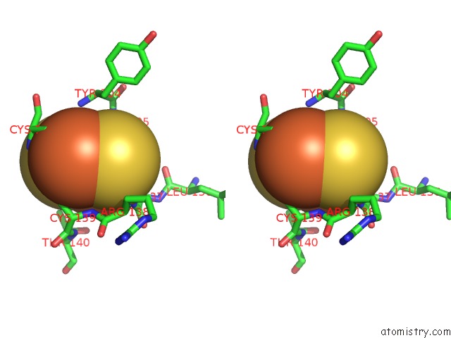
 Stereo pair view
|
A full contact list of Iron with other atoms in the Fe binding
site number 2 of Crystal Structure of the Cu,Mo-Co Dehydrogenase (Codh); Cyanide- Inactivated Form within 5.0Å range:
probe
|
atom
|
residue
|
distance (Å)
|
B
|
Occ
|
A:Fe4907
b:9.9
occ:1.00
|
FE2
|
A:FES4907
|
0.0
|
9.9
|
1.0
|
S1
|
A:FES4907
|
2.2
|
9.7
|
1.0
|
S2
|
A:FES4907
|
2.2
|
9.7
|
1.0
|
SG
|
A:CYS105
|
2.3
|
9.9
|
1.0
|
SG
|
A:CYS137
|
2.3
|
9.3
|
1.0
|
FE1
|
A:FES4907
|
2.7
|
10.0
|
1.0
|
CB
|
A:CYS105
|
3.3
|
10.5
|
1.0
|
CB
|
A:CYS137
|
3.4
|
10.2
|
1.0
|
CA
|
A:CYS137
|
3.7
|
10.0
|
1.0
|
N
|
A:CYS105
|
4.0
|
9.5
|
1.0
|
N
|
A:ARG138
|
4.1
|
9.6
|
1.0
|
N
|
A:CYS139
|
4.2
|
9.9
|
1.0
|
CA
|
A:CYS105
|
4.3
|
10.2
|
1.0
|
C
|
A:CYS137
|
4.3
|
9.7
|
1.0
|
O
|
A:HOH4912
|
4.3
|
11.6
|
1.0
|
CB
|
A:CYS139
|
4.4
|
10.7
|
1.0
|
SG
|
A:CYS139
|
4.4
|
9.7
|
1.0
|
SG
|
A:CYS102
|
4.7
|
9.2
|
1.0
|
CG2
|
A:THR140
|
4.7
|
10.4
|
1.0
|
CA
|
A:CYS139
|
4.9
|
10.4
|
1.0
|
N
|
A:TYR104
|
4.9
|
9.9
|
1.0
|
C
|
A:CYS105
|
4.9
|
10.2
|
1.0
|
O
|
A:LEU136
|
4.9
|
11.6
|
1.0
|
N
|
A:CYS137
|
5.0
|
10.3
|
1.0
|
|
Iron binding site 3 out
of 8 in 1n60
Go back to
Iron Binding Sites List in 1n60
Iron binding site 3 out
of 8 in the Crystal Structure of the Cu,Mo-Co Dehydrogenase (Codh); Cyanide- Inactivated Form
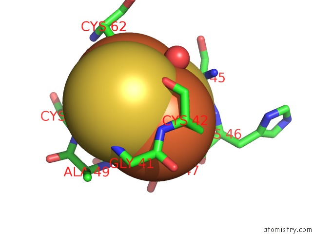
 Mono view
 Stereo pair view
|
A full contact list of Iron with other atoms in the Fe binding
site number 3 of Crystal Structure of the Cu,Mo-Co Dehydrogenase (Codh); Cyanide- Inactivated Form within 5.0Å range:
probe
|
atom
|
residue
|
distance (Å)
|
B
|
Occ
|
A:Fe4908
b:10.4
occ:1.00
|
FE1
|
A:FES4908
|
0.0
|
10.4
|
1.0
|
S2
|
A:FES4908
|
2.2
|
10.0
|
1.0
|
S1
|
A:FES4908
|
2.3
|
9.9
|
1.0
|
SG
|
A:CYS47
|
2.3
|
9.9
|
1.0
|
SG
|
A:CYS42
|
2.3
|
10.3
|
1.0
|
FE2
|
A:FES4908
|
2.7
|
10.4
|
1.0
|
CB
|
A:CYS47
|
3.4
|
11.3
|
1.0
|
N
|
A:CYS47
|
3.4
|
10.2
|
1.0
|
CB
|
A:CYS42
|
3.4
|
11.5
|
1.0
|
N
|
A:CYS42
|
3.5
|
11.5
|
1.0
|
CA
|
A:CYS47
|
3.8
|
11.2
|
1.0
|
N
|
A:GLY48
|
3.8
|
9.9
|
1.0
|
CA
|
A:CYS42
|
3.9
|
11.1
|
1.0
|
O
|
A:HOH4953
|
3.9
|
18.0
|
1.0
|
O
|
A:CYS42
|
4.0
|
12.7
|
1.0
|
N
|
A:GLY41
|
4.1
|
11.3
|
1.0
|
C
|
A:CYS42
|
4.1
|
11.3
|
1.0
|
C
|
A:GLY41
|
4.2
|
11.0
|
1.0
|
C
|
A:CYS47
|
4.2
|
10.4
|
1.0
|
SG
|
A:CYS62
|
4.3
|
10.5
|
1.0
|
N
|
A:HIS46
|
4.4
|
10.6
|
1.0
|
CA
|
A:GLY41
|
4.4
|
11.5
|
1.0
|
N
|
A:ALA49
|
4.5
|
10.0
|
1.0
|
C
|
A:HIS46
|
4.5
|
10.4
|
1.0
|
CA
|
A:SER45
|
4.6
|
11.2
|
1.0
|
SG
|
A:CYS50
|
4.6
|
10.0
|
1.0
|
C
|
A:SER45
|
4.6
|
10.5
|
1.0
|
N
|
A:SER45
|
4.7
|
10.6
|
1.0
|
CA
|
A:GLY48
|
4.8
|
10.6
|
1.0
|
O
|
A:GLY41
|
4.9
|
12.7
|
1.0
|
|
Iron binding site 4 out
of 8 in 1n60
Go back to
Iron Binding Sites List in 1n60
Iron binding site 4 out
of 8 in the Crystal Structure of the Cu,Mo-Co Dehydrogenase (Codh); Cyanide- Inactivated Form
 Mono view
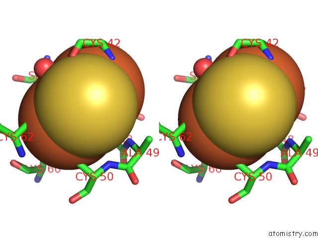
 Stereo pair view
|
A full contact list of Iron with other atoms in the Fe binding
site number 4 of Crystal Structure of the Cu,Mo-Co Dehydrogenase (Codh); Cyanide- Inactivated Form within 5.0Å range:
probe
|
atom
|
residue
|
distance (Å)
|
B
|
Occ
|
A:Fe4908
b:10.4
occ:1.00
|
FE2
|
A:FES4908
|
0.0
|
10.4
|
1.0
|
S1
|
A:FES4908
|
2.2
|
9.9
|
1.0
|
S2
|
A:FES4908
|
2.2
|
10.0
|
1.0
|
SG
|
A:CYS50
|
2.3
|
10.0
|
1.0
|
SG
|
A:CYS62
|
2.3
|
10.5
|
1.0
|
FE1
|
A:FES4908
|
2.7
|
10.4
|
1.0
|
CB
|
A:CYS62
|
3.2
|
11.4
|
1.0
|
CB
|
A:CYS50
|
3.4
|
10.8
|
1.0
|
O
|
A:HOH4953
|
4.0
|
18.0
|
1.0
|
N
|
A:CYS62
|
4.2
|
10.9
|
1.0
|
N
|
A:CYS50
|
4.3
|
10.2
|
1.0
|
CA
|
A:CYS62
|
4.3
|
11.5
|
1.0
|
CB
|
A:LYS60
|
4.4
|
11.0
|
1.0
|
SG
|
A:CYS47
|
4.4
|
9.9
|
1.0
|
CA
|
A:CYS50
|
4.5
|
10.6
|
1.0
|
SG
|
A:CYS42
|
4.5
|
10.3
|
1.0
|
N
|
A:GLY48
|
4.6
|
9.9
|
1.0
|
CG
|
A:LYS60
|
4.8
|
10.9
|
1.0
|
N
|
A:ALA49
|
4.8
|
10.0
|
1.0
|
CA
|
A:SER45
|
4.8
|
11.2
|
1.0
|
CA
|
A:GLY48
|
4.9
|
10.6
|
1.0
|
CA
|
A:LYS60
|
5.0
|
9.8
|
1.0
|
|
Iron binding site 5 out
of 8 in 1n60
Go back to
Iron Binding Sites List in 1n60
Iron binding site 5 out
of 8 in the Crystal Structure of the Cu,Mo-Co Dehydrogenase (Codh); Cyanide- Inactivated Form
 Mono view
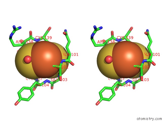
 Stereo pair view
|
A full contact list of Iron with other atoms in the Fe binding
site number 5 of Crystal Structure of the Cu,Mo-Co Dehydrogenase (Codh); Cyanide- Inactivated Form within 5.0Å range:
probe
|
atom
|
residue
|
distance (Å)
|
B
|
Occ
|
D:Fe5907
b:10.2
occ:1.00
|
FE1
|
D:FES5907
|
0.0
|
10.2
|
1.0
|
S2
|
D:FES5907
|
2.3
|
9.9
|
1.0
|
S1
|
D:FES5907
|
2.3
|
10.2
|
1.0
|
SG
|
D:CYS102
|
2.3
|
9.7
|
1.0
|
SG
|
D:CYS139
|
2.3
|
9.9
|
1.0
|
FE2
|
D:FES5907
|
2.7
|
10.3
|
1.0
|
CB
|
D:CYS139
|
3.3
|
11.0
|
1.0
|
CB
|
D:CYS102
|
3.4
|
11.2
|
1.0
|
N
|
D:CYS102
|
3.6
|
10.2
|
1.0
|
CA
|
D:CYS102
|
3.9
|
10.5
|
1.0
|
O
|
E:HOH5930
|
3.9
|
11.5
|
1.0
|
N
|
D:GLY103
|
4.0
|
10.3
|
1.0
|
N
|
D:CYS139
|
4.0
|
10.5
|
1.0
|
N
|
D:TYR104
|
4.1
|
10.9
|
1.0
|
CA
|
D:CYS139
|
4.2
|
10.6
|
1.0
|
C
|
D:CYS102
|
4.3
|
10.8
|
1.0
|
SG
|
D:CYS137
|
4.4
|
9.6
|
1.0
|
C
|
D:GLN101
|
4.7
|
10.1
|
1.0
|
N
|
D:CYS105
|
4.7
|
10.2
|
1.0
|
SG
|
D:CYS105
|
4.7
|
10.0
|
1.0
|
CB
|
D:TYR104
|
4.8
|
10.8
|
1.0
|
N
|
D:ARG138
|
4.8
|
10.3
|
1.0
|
N
|
D:GLN101
|
4.8
|
10.1
|
1.0
|
CA
|
D:GLY103
|
4.9
|
10.5
|
1.0
|
C
|
D:ARG138
|
4.9
|
10.0
|
1.0
|
CA
|
D:TYR104
|
4.9
|
10.4
|
1.0
|
CB
|
D:GLN101
|
4.9
|
10.5
|
1.0
|
C
|
D:GLY103
|
5.0
|
10.2
|
1.0
|
|
Iron binding site 6 out
of 8 in 1n60
Go back to
Iron Binding Sites List in 1n60
Iron binding site 6 out
of 8 in the Crystal Structure of the Cu,Mo-Co Dehydrogenase (Codh); Cyanide- Inactivated Form
 Mono view
 Stereo pair view
|
A full contact list of Iron with other atoms in the Fe binding
site number 6 of Crystal Structure of the Cu,Mo-Co Dehydrogenase (Codh); Cyanide- Inactivated Form within 5.0Å range:
probe
|
atom
|
residue
|
distance (Å)
|
B
|
Occ
|
D:Fe5907
b:10.3
occ:1.00
|
FE2
|
D:FES5907
|
0.0
|
10.3
|
1.0
|
S1
|
D:FES5907
|
2.2
|
10.2
|
1.0
|
S2
|
D:FES5907
|
2.2
|
9.9
|
1.0
|
SG
|
D:CYS105
|
2.3
|
10.0
|
1.0
|
SG
|
D:CYS137
|
2.3
|
9.6
|
1.0
|
FE1
|
D:FES5907
|
2.7
|
10.2
|
1.0
|
CB
|
D:CYS105
|
3.3
|
10.3
|
1.0
|
CB
|
D:CYS137
|
3.4
|
11.3
|
1.0
|
CA
|
D:CYS137
|
3.7
|
10.3
|
1.0
|
N
|
D:CYS105
|
4.0
|
10.2
|
1.0
|
N
|
D:ARG138
|
4.1
|
10.3
|
1.0
|
CA
|
D:CYS105
|
4.2
|
9.9
|
1.0
|
N
|
D:CYS139
|
4.2
|
10.5
|
1.0
|
O
|
D:HOH5916
|
4.3
|
11.8
|
1.0
|
CB
|
D:CYS139
|
4.4
|
11.0
|
1.0
|
C
|
D:CYS137
|
4.4
|
9.8
|
1.0
|
SG
|
D:CYS139
|
4.4
|
9.9
|
1.0
|
CG2
|
D:THR140
|
4.7
|
11.2
|
1.0
|
SG
|
D:CYS102
|
4.7
|
9.7
|
1.0
|
CA
|
D:CYS139
|
4.9
|
10.6
|
1.0
|
C
|
D:CYS105
|
4.9
|
10.3
|
1.0
|
N
|
D:TYR104
|
4.9
|
10.9
|
1.0
|
O
|
D:LEU136
|
5.0
|
11.5
|
1.0
|
N
|
D:THR140
|
5.0
|
10.0
|
1.0
|
|
Iron binding site 7 out
of 8 in 1n60
Go back to
Iron Binding Sites List in 1n60
Iron binding site 7 out
of 8 in the Crystal Structure of the Cu,Mo-Co Dehydrogenase (Codh); Cyanide- Inactivated Form
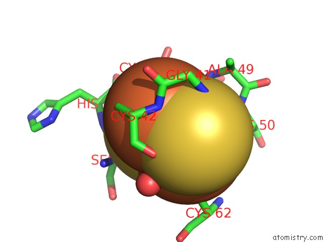
 Mono view
 Stereo pair view
|
A full contact list of Iron with other atoms in the Fe binding
site number 7 of Crystal Structure of the Cu,Mo-Co Dehydrogenase (Codh); Cyanide- Inactivated Form within 5.0Å range:
probe
|
atom
|
residue
|
distance (Å)
|
B
|
Occ
|
D:Fe5908
b:10.6
occ:1.00
|
FE1
|
D:FES5908
|
0.0
|
10.6
|
1.0
|
S2
|
D:FES5908
|
2.2
|
10.3
|
1.0
|
S1
|
D:FES5908
|
2.3
|
10.1
|
1.0
|
SG
|
D:CYS47
|
2.3
|
10.0
|
1.0
|
SG
|
D:CYS42
|
2.3
|
10.2
|
1.0
|
FE2
|
D:FES5908
|
2.7
|
10.6
|
1.0
|
CB
|
D:CYS47
|
3.4
|
10.8
|
1.0
|
N
|
D:CYS47
|
3.4
|
10.1
|
1.0
|
CB
|
D:CYS42
|
3.5
|
11.3
|
1.0
|
N
|
D:CYS42
|
3.5
|
11.1
|
1.0
|
CA
|
D:CYS47
|
3.8
|
11.1
|
1.0
|
CA
|
D:CYS42
|
3.9
|
11.1
|
1.0
|
N
|
D:GLY48
|
3.9
|
10.7
|
1.0
|
O
|
D:CYS42
|
4.0
|
12.2
|
1.0
|
C
|
D:CYS42
|
4.1
|
11.0
|
1.0
|
C
|
D:GLY41
|
4.2
|
11.0
|
1.0
|
N
|
D:GLY41
|
4.2
|
10.8
|
1.0
|
C
|
D:CYS47
|
4.2
|
10.9
|
1.0
|
O
|
F:HOH5962
|
4.2
|
19.6
|
1.0
|
SG
|
D:CYS62
|
4.3
|
10.2
|
1.0
|
N
|
D:HIS46
|
4.4
|
11.3
|
1.0
|
CA
|
D:GLY41
|
4.5
|
11.2
|
1.0
|
N
|
D:ALA49
|
4.5
|
10.1
|
1.0
|
C
|
D:HIS46
|
4.6
|
9.9
|
1.0
|
SG
|
D:CYS50
|
4.6
|
10.2
|
1.0
|
CA
|
D:SER45
|
4.6
|
10.5
|
1.0
|
C
|
D:SER45
|
4.7
|
10.7
|
1.0
|
N
|
D:SER45
|
4.7
|
11.4
|
1.0
|
CA
|
D:GLY48
|
4.8
|
10.0
|
1.0
|
O
|
D:GLY41
|
4.9
|
12.2
|
1.0
|
|
Iron binding site 8 out
of 8 in 1n60
Go back to
Iron Binding Sites List in 1n60
Iron binding site 8 out
of 8 in the Crystal Structure of the Cu,Mo-Co Dehydrogenase (Codh); Cyanide- Inactivated Form
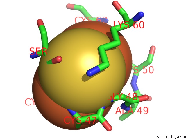
 Mono view
 Stereo pair view
|
A full contact list of Iron with other atoms in the Fe binding
site number 8 of Crystal Structure of the Cu,Mo-Co Dehydrogenase (Codh); Cyanide- Inactivated Form within 5.0Å range:
probe
|
atom
|
residue
|
distance (Å)
|
B
|
Occ
|
D:Fe5908
b:10.6
occ:1.00
|
FE2
|
D:FES5908
|
0.0
|
10.6
|
1.0
|
S1
|
D:FES5908
|
2.2
|
10.1
|
1.0
|
S2
|
D:FES5908
|
2.2
|
10.3
|
1.0
|
SG
|
D:CYS50
|
2.3
|
10.2
|
1.0
|
SG
|
D:CYS62
|
2.3
|
10.2
|
1.0
|
FE1
|
D:FES5908
|
2.7
|
10.6
|
1.0
|
CB
|
D:CYS62
|
3.2
|
11.4
|
1.0
|
CB
|
D:CYS50
|
3.4
|
10.6
|
1.0
|
O
|
F:HOH5962
|
4.1
|
19.6
|
1.0
|
N
|
D:CYS62
|
4.2
|
10.7
|
1.0
|
N
|
D:CYS50
|
4.3
|
10.4
|
1.0
|
CA
|
D:CYS62
|
4.3
|
11.1
|
1.0
|
SG
|
D:CYS47
|
4.4
|
10.0
|
1.0
|
CB
|
D:LYS60
|
4.4
|
12.0
|
1.0
|
CA
|
D:CYS50
|
4.5
|
10.9
|
1.0
|
SG
|
D:CYS42
|
4.5
|
10.2
|
1.0
|
N
|
D:GLY48
|
4.6
|
10.7
|
1.0
|
CA
|
D:SER45
|
4.8
|
10.5
|
1.0
|
N
|
D:ALA49
|
4.8
|
10.1
|
1.0
|
CG
|
D:LYS60
|
4.8
|
11.0
|
1.0
|
CA
|
D:GLY48
|
4.9
|
10.0
|
1.0
|
CA
|
D:LYS60
|
5.0
|
11.3
|
1.0
|
|
Reference:
H.Dobbek,
L.Gremer,
R.Kiefersauer,
R.Huber,
O.Meyer.
Catalysis at A Dinuclear [Cusmo(=O)Oh] Cluster in A Co Dehydrogenase Resolved at 1.1-A Resolution Proc.Natl.Acad.Sci.Usa V. 99 15971 2002.
ISSN: ISSN 0027-8424
PubMed: 12475995
DOI: 10.1073/PNAS.212640899
Page generated: Sat Aug 3 11:25:13 2024
|