Iron »
PDB 1n5w-1nmi »
1n62 »
Iron in PDB 1n62: Crystal Structure of the Mo,Cu-Co Dehydrogenase (Codh), N- Butylisocyanide-Bound State
Enzymatic activity of Crystal Structure of the Mo,Cu-Co Dehydrogenase (Codh), N- Butylisocyanide-Bound State
All present enzymatic activity of Crystal Structure of the Mo,Cu-Co Dehydrogenase (Codh), N- Butylisocyanide-Bound State:
1.2.99.2;
1.2.99.2;
Protein crystallography data
The structure of Crystal Structure of the Mo,Cu-Co Dehydrogenase (Codh), N- Butylisocyanide-Bound State, PDB code: 1n62
was solved by
H.Dobbek,
L.Gremer,
R.Kiefersauer,
R.Huber,
O.Meyer,
with X-Ray Crystallography technique. A brief refinement statistics is given in the table below:
| Resolution Low / High (Å) | 17.80 / 1.09 |
| Space group | P 21 21 21 |
| Cell size a, b, c (Å), α, β, γ (°) | 117.860, 130.019, 156.231, 90.00, 90.00, 90.00 |
| R / Rfree (%) | 14.4 / 17.2 |
Other elements in 1n62:
The structure of Crystal Structure of the Mo,Cu-Co Dehydrogenase (Codh), N- Butylisocyanide-Bound State also contains other interesting chemical elements:
| Molybdenum | (Mo) | 2 atoms |
| Copper | (Cu) | 2 atoms |
Iron Binding Sites:
The binding sites of Iron atom in the Crystal Structure of the Mo,Cu-Co Dehydrogenase (Codh), N- Butylisocyanide-Bound State
(pdb code 1n62). This binding sites where shown within
5.0 Angstroms radius around Iron atom.
In total 8 binding sites of Iron where determined in the Crystal Structure of the Mo,Cu-Co Dehydrogenase (Codh), N- Butylisocyanide-Bound State, PDB code: 1n62:
Jump to Iron binding site number: 1; 2; 3; 4; 5; 6; 7; 8;
In total 8 binding sites of Iron where determined in the Crystal Structure of the Mo,Cu-Co Dehydrogenase (Codh), N- Butylisocyanide-Bound State, PDB code: 1n62:
Jump to Iron binding site number: 1; 2; 3; 4; 5; 6; 7; 8;
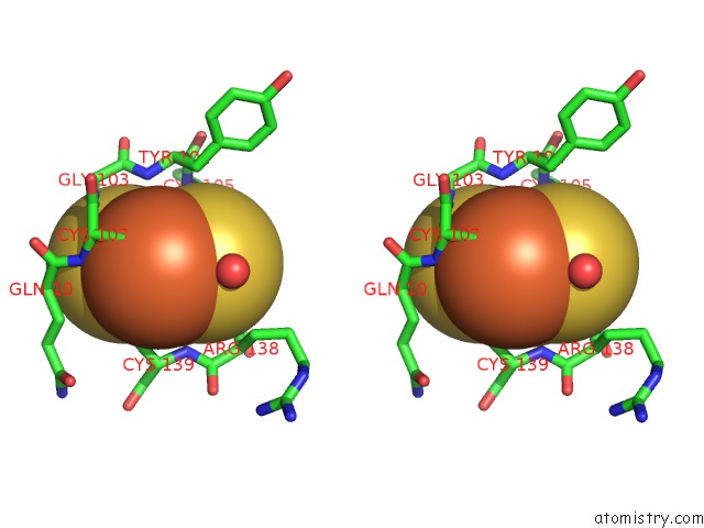
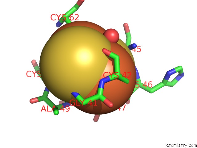
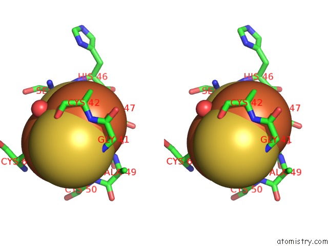
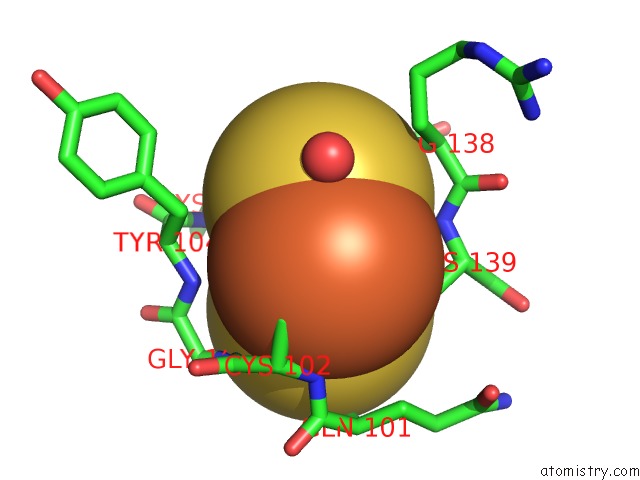
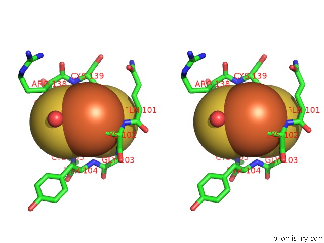
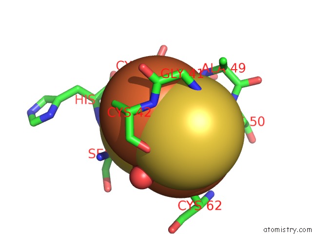
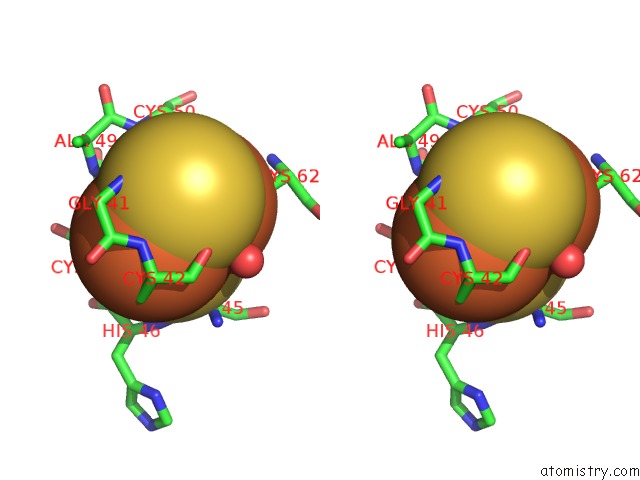
Iron binding site 1 out of 8 in 1n62
Go back to
Iron binding site 1 out
of 8 in the Crystal Structure of the Mo,Cu-Co Dehydrogenase (Codh), N- Butylisocyanide-Bound State
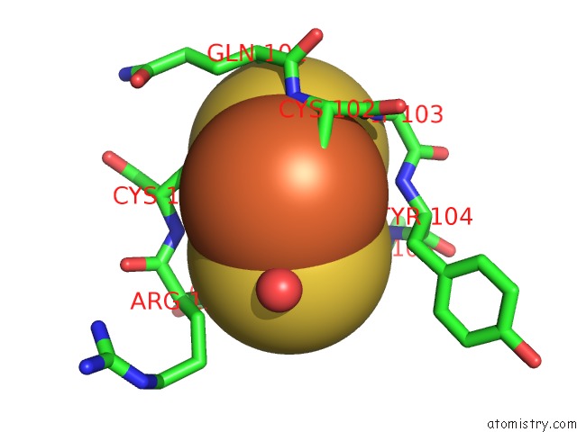
Mono view
Stereo pair view
Mono view
Stereo pair view
A full contact list of Iron with other atoms in the Fe binding
site number 1 of Crystal Structure of the Mo,Cu-Co Dehydrogenase (Codh), N- Butylisocyanide-Bound State within 5.0Å range:
|
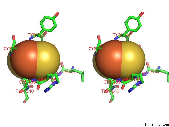
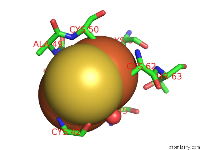
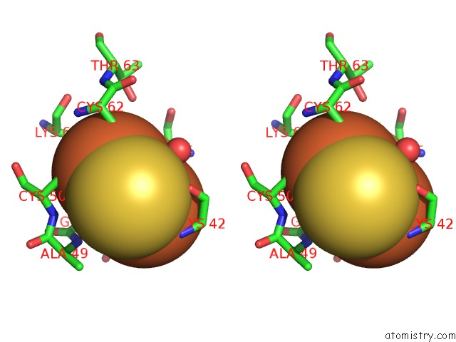
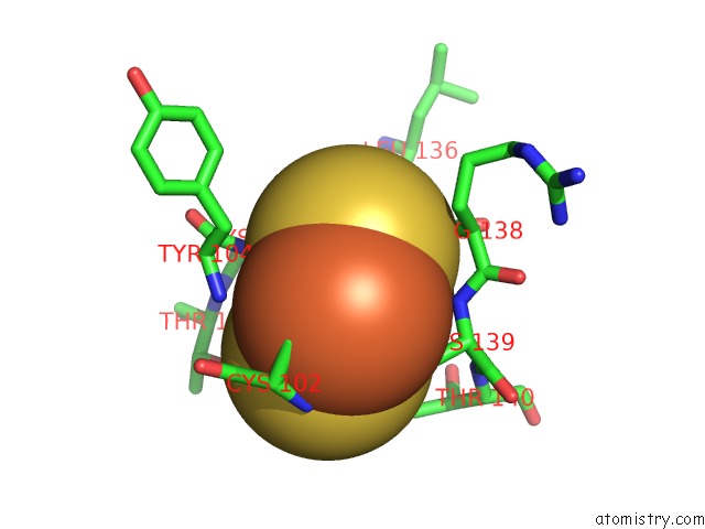
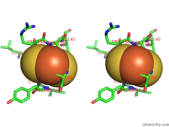
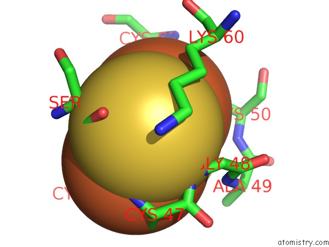
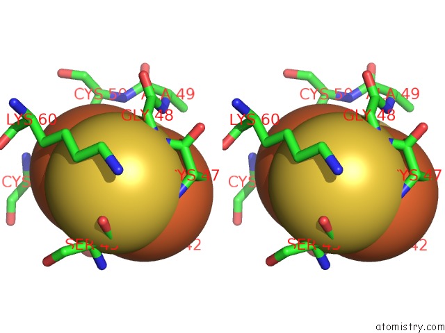
Iron binding site 2 out of 8 in 1n62
Go back to
Iron binding site 2 out
of 8 in the Crystal Structure of the Mo,Cu-Co Dehydrogenase (Codh), N- Butylisocyanide-Bound State
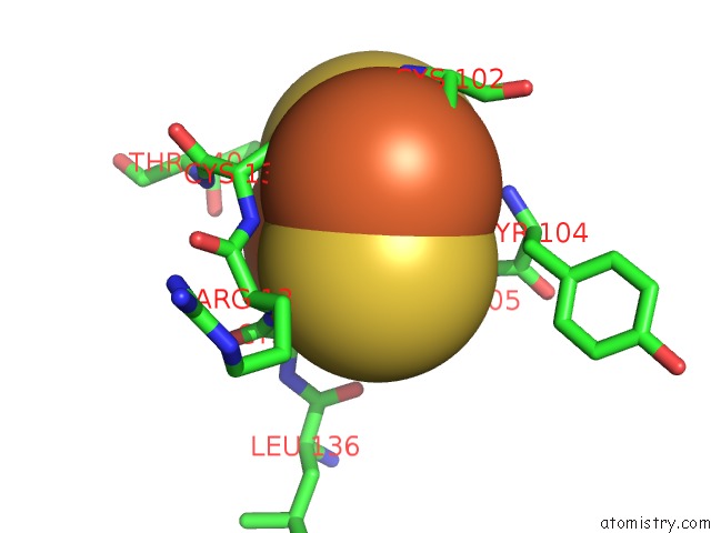
Mono view
Stereo pair view
Mono view
Stereo pair view
A full contact list of Iron with other atoms in the Fe binding
site number 2 of Crystal Structure of the Mo,Cu-Co Dehydrogenase (Codh), N- Butylisocyanide-Bound State within 5.0Å range:
|
Iron binding site 3 out of 8 in 1n62
Go back to
Iron binding site 3 out
of 8 in the Crystal Structure of the Mo,Cu-Co Dehydrogenase (Codh), N- Butylisocyanide-Bound State
Mono view
Stereo pair view
Mono view
Stereo pair view
A full contact list of Iron with other atoms in the Fe binding
site number 3 of Crystal Structure of the Mo,Cu-Co Dehydrogenase (Codh), N- Butylisocyanide-Bound State within 5.0Å range:
|
Iron binding site 4 out of 8 in 1n62
Go back to
Iron binding site 4 out
of 8 in the Crystal Structure of the Mo,Cu-Co Dehydrogenase (Codh), N- Butylisocyanide-Bound State
Mono view
Stereo pair view
Mono view
Stereo pair view
A full contact list of Iron with other atoms in the Fe binding
site number 4 of Crystal Structure of the Mo,Cu-Co Dehydrogenase (Codh), N- Butylisocyanide-Bound State within 5.0Å range:
|
Iron binding site 5 out of 8 in 1n62
Go back to
Iron binding site 5 out
of 8 in the Crystal Structure of the Mo,Cu-Co Dehydrogenase (Codh), N- Butylisocyanide-Bound State
Mono view
Stereo pair view
Mono view
Stereo pair view
A full contact list of Iron with other atoms in the Fe binding
site number 5 of Crystal Structure of the Mo,Cu-Co Dehydrogenase (Codh), N- Butylisocyanide-Bound State within 5.0Å range:
|
Iron binding site 6 out of 8 in 1n62
Go back to
Iron binding site 6 out
of 8 in the Crystal Structure of the Mo,Cu-Co Dehydrogenase (Codh), N- Butylisocyanide-Bound State
Mono view
Stereo pair view
Mono view
Stereo pair view
A full contact list of Iron with other atoms in the Fe binding
site number 6 of Crystal Structure of the Mo,Cu-Co Dehydrogenase (Codh), N- Butylisocyanide-Bound State within 5.0Å range:
|
Iron binding site 7 out of 8 in 1n62
Go back to
Iron binding site 7 out
of 8 in the Crystal Structure of the Mo,Cu-Co Dehydrogenase (Codh), N- Butylisocyanide-Bound State
Mono view
Stereo pair view
Mono view
Stereo pair view
A full contact list of Iron with other atoms in the Fe binding
site number 7 of Crystal Structure of the Mo,Cu-Co Dehydrogenase (Codh), N- Butylisocyanide-Bound State within 5.0Å range:
|
Iron binding site 8 out of 8 in 1n62
Go back to
Iron binding site 8 out
of 8 in the Crystal Structure of the Mo,Cu-Co Dehydrogenase (Codh), N- Butylisocyanide-Bound State
Mono view
Stereo pair view
Mono view
Stereo pair view
A full contact list of Iron with other atoms in the Fe binding
site number 8 of Crystal Structure of the Mo,Cu-Co Dehydrogenase (Codh), N- Butylisocyanide-Bound State within 5.0Å range:
|
Reference:
H.Dobbek,
L.Gremer,
R.Kiefersauer,
R.Huber,
O.Meyer.
Catalysis at A Dinuclear [Cusmo(=O)Oh] Cluster in A Co Dehydrogenase Resolved at 1.1-A Resolution Proc.Natl.Acad.Sci.Usa V. 99 15971 2002.
ISSN: ISSN 0027-8424
PubMed: 12475995
DOI: 10.1073/PNAS.212640899
Page generated: Wed Jul 16 18:30:35 2025
ISSN: ISSN 0027-8424
PubMed: 12475995
DOI: 10.1073/PNAS.212640899
Last articles
Fe in 2YXOFe in 2YRS
Fe in 2YXC
Fe in 2YNM
Fe in 2YVJ
Fe in 2YP1
Fe in 2YU2
Fe in 2YU1
Fe in 2YQB
Fe in 2YOO