Iron »
PDB 1n5w-1nmi »
1n6b »
Iron in PDB 1n6b: Microsomal Cytochrome P450 2C5/3LVDH Complex with A Dimethyl Derivative of Sulfaphenazole
Enzymatic activity of Microsomal Cytochrome P450 2C5/3LVDH Complex with A Dimethyl Derivative of Sulfaphenazole
All present enzymatic activity of Microsomal Cytochrome P450 2C5/3LVDH Complex with A Dimethyl Derivative of Sulfaphenazole:
1.14.14.1;
1.14.14.1;
Protein crystallography data
The structure of Microsomal Cytochrome P450 2C5/3LVDH Complex with A Dimethyl Derivative of Sulfaphenazole, PDB code: 1n6b
was solved by
M.R.Wester,
E.F.Johnson,
C.Marques-Soares,
P.M.Dansette,
D.Mansuy,
C.D.Stout,
with X-Ray Crystallography technique. A brief refinement statistics is given in the table below:
| Resolution Low / High (Å) | 50.00 / 2.30 |
| Space group | I 2 2 2 |
| Cell size a, b, c (Å), α, β, γ (°) | 74.330, 134.290, 171.840, 90.00, 90.00, 90.00 |
| R / Rfree (%) | 25.7 / 29.2 |
Iron Binding Sites:
The binding sites of Iron atom in the Microsomal Cytochrome P450 2C5/3LVDH Complex with A Dimethyl Derivative of Sulfaphenazole
(pdb code 1n6b). This binding sites where shown within
5.0 Angstroms radius around Iron atom.
In total only one binding site of Iron was determined in the Microsomal Cytochrome P450 2C5/3LVDH Complex with A Dimethyl Derivative of Sulfaphenazole, PDB code: 1n6b:
In total only one binding site of Iron was determined in the Microsomal Cytochrome P450 2C5/3LVDH Complex with A Dimethyl Derivative of Sulfaphenazole, PDB code: 1n6b:
Iron binding site 1 out of 1 in 1n6b
Go back to
Iron binding site 1 out
of 1 in the Microsomal Cytochrome P450 2C5/3LVDH Complex with A Dimethyl Derivative of Sulfaphenazole
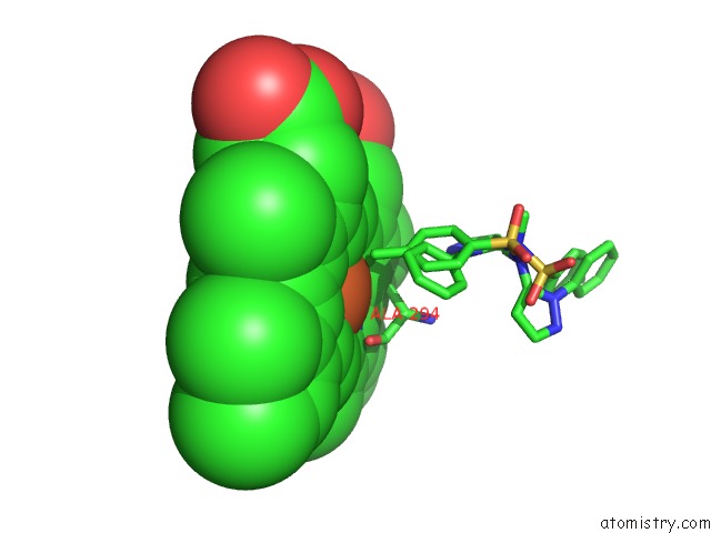
Mono view
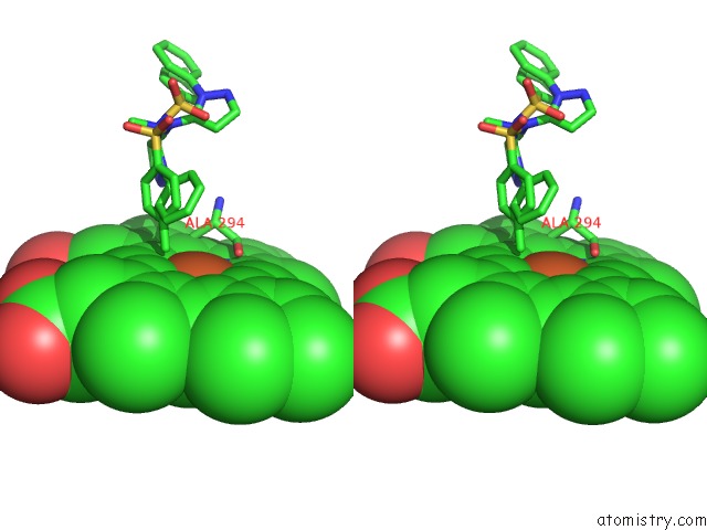
Stereo pair view
Mono view
Stereo pair view
A full contact list of Iron with other atoms in the Fe binding
site number 1 of Microsomal Cytochrome P450 2C5/3LVDH Complex with A Dimethyl Derivative of Sulfaphenazole within 5.0Å range:
|
Reference:
M.R.Wester,
E.F.Johnson,
C.Marques-Soares,
P.M.Dansette,
D.Mansuy,
C.D.Stout.
Structure of A Substrate Complex of Mammalian Cytochrome P450 2C5 at 2.3 A Resolution: Evidence For Multiple Substrate Binding Modes Biochemistry V. 42 6370 2003.
ISSN: ISSN 0006-2960
PubMed: 12767218
DOI: 10.1021/BI0273922
Page generated: Sat Aug 3 11:25:12 2024
ISSN: ISSN 0006-2960
PubMed: 12767218
DOI: 10.1021/BI0273922
Last articles
F in 4QJ3F in 4QAB
F in 4QIY
F in 4QIZ
F in 4QIN
F in 4QGH
F in 4QGG
F in 4QGA
F in 4Q0L
F in 4QBX