Iron »
PDB 1n5w-1nmi »
1nej »
Iron in PDB 1nej: Crystalline Human Carbonmonoxy Hemoglobin S (Liganded Sickle Cell Hemoglobin) Exhibits the R2 Quaternary State at Neutral pH in the Presence of Polyethylene Glycol: the 2.1 Angstrom Resolution Crystal Structure
Protein crystallography data
The structure of Crystalline Human Carbonmonoxy Hemoglobin S (Liganded Sickle Cell Hemoglobin) Exhibits the R2 Quaternary State at Neutral pH in the Presence of Polyethylene Glycol: the 2.1 Angstrom Resolution Crystal Structure, PDB code: 1nej
was solved by
L.N.Patskovska,
Y.V.Patskovsky,
S.C.Almo,
R.E.Hirsch,
with X-Ray Crystallography technique. A brief refinement statistics is given in the table below:
| Resolution Low / High (Å) | 8.00 / 2.10 |
| Space group | P 21 21 21 |
| Cell size a, b, c (Å), α, β, γ (°) | 58.000, 58.530, 171.840, 90.00, 90.00, 90.00 |
| R / Rfree (%) | 23.5 / 26.8 |
Iron Binding Sites:
The binding sites of Iron atom in the Crystalline Human Carbonmonoxy Hemoglobin S (Liganded Sickle Cell Hemoglobin) Exhibits the R2 Quaternary State at Neutral pH in the Presence of Polyethylene Glycol: the 2.1 Angstrom Resolution Crystal Structure
(pdb code 1nej). This binding sites where shown within
5.0 Angstroms radius around Iron atom.
In total 4 binding sites of Iron where determined in the Crystalline Human Carbonmonoxy Hemoglobin S (Liganded Sickle Cell Hemoglobin) Exhibits the R2 Quaternary State at Neutral pH in the Presence of Polyethylene Glycol: the 2.1 Angstrom Resolution Crystal Structure, PDB code: 1nej:
Jump to Iron binding site number: 1; 2; 3; 4;
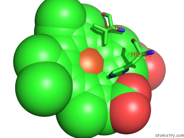
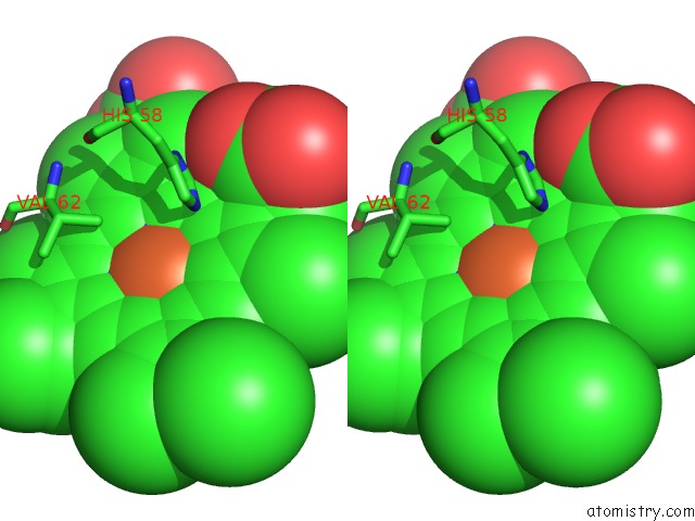
Iron binding site 1 out of 4 in 1nej
Go back to
Iron binding site 1 out
of 4 in the Crystalline Human Carbonmonoxy Hemoglobin S (Liganded Sickle Cell Hemoglobin) Exhibits the R2 Quaternary State at Neutral pH in the Presence of Polyethylene Glycol: the 2.1 Angstrom Resolution Crystal Structure
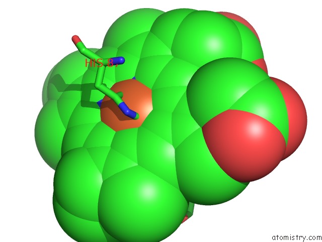
Mono view
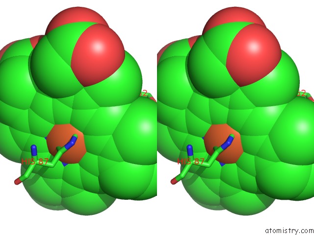
Stereo pair view
Mono view
Stereo pair view
A full contact list of Iron with other atoms in the Fe binding
site number 1 of Crystalline Human Carbonmonoxy Hemoglobin S (Liganded Sickle Cell Hemoglobin) Exhibits the R2 Quaternary State at Neutral pH in the Presence of Polyethylene Glycol: the 2.1 Angstrom Resolution Crystal Structure within 5.0Å range:
|
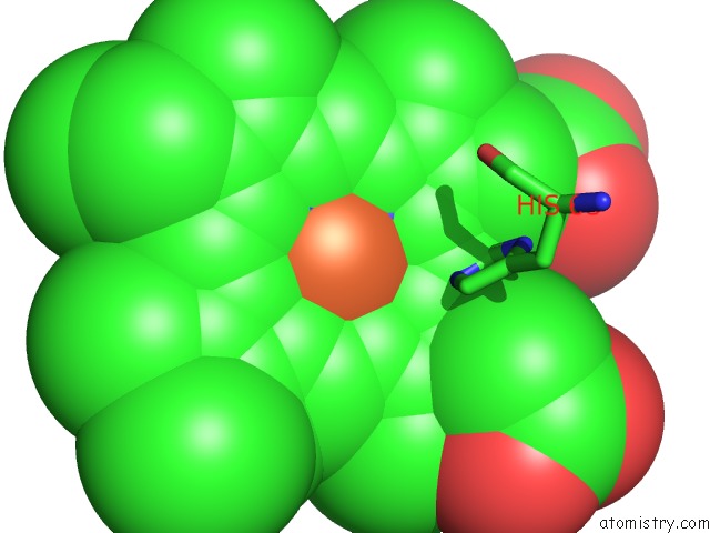
Iron binding site 2 out of 4 in 1nej
Go back to
Iron binding site 2 out
of 4 in the Crystalline Human Carbonmonoxy Hemoglobin S (Liganded Sickle Cell Hemoglobin) Exhibits the R2 Quaternary State at Neutral pH in the Presence of Polyethylene Glycol: the 2.1 Angstrom Resolution Crystal Structure
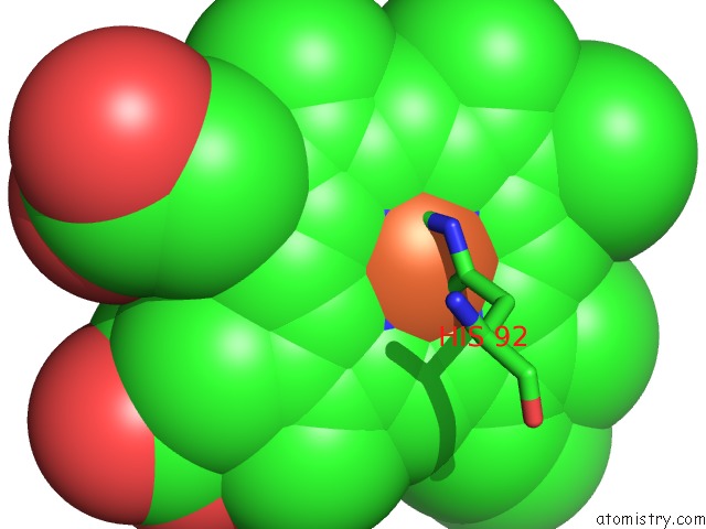
Mono view
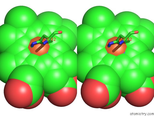
Stereo pair view
Mono view
Stereo pair view
A full contact list of Iron with other atoms in the Fe binding
site number 2 of Crystalline Human Carbonmonoxy Hemoglobin S (Liganded Sickle Cell Hemoglobin) Exhibits the R2 Quaternary State at Neutral pH in the Presence of Polyethylene Glycol: the 2.1 Angstrom Resolution Crystal Structure within 5.0Å range:
|
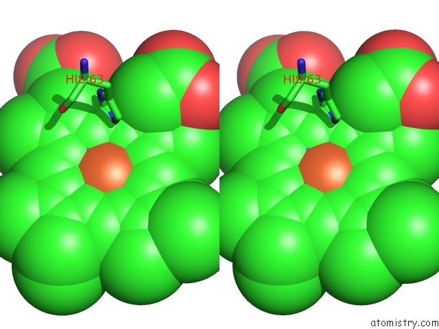
Iron binding site 3 out of 4 in 1nej
Go back to
Iron binding site 3 out
of 4 in the Crystalline Human Carbonmonoxy Hemoglobin S (Liganded Sickle Cell Hemoglobin) Exhibits the R2 Quaternary State at Neutral pH in the Presence of Polyethylene Glycol: the 2.1 Angstrom Resolution Crystal Structure
Mono view
Stereo pair view
Mono view
Stereo pair view
A full contact list of Iron with other atoms in the Fe binding
site number 3 of Crystalline Human Carbonmonoxy Hemoglobin S (Liganded Sickle Cell Hemoglobin) Exhibits the R2 Quaternary State at Neutral pH in the Presence of Polyethylene Glycol: the 2.1 Angstrom Resolution Crystal Structure within 5.0Å range:
|
Iron binding site 4 out of 4 in 1nej
Go back to
Iron binding site 4 out
of 4 in the Crystalline Human Carbonmonoxy Hemoglobin S (Liganded Sickle Cell Hemoglobin) Exhibits the R2 Quaternary State at Neutral pH in the Presence of Polyethylene Glycol: the 2.1 Angstrom Resolution Crystal Structure
Mono view
Stereo pair view
Mono view
Stereo pair view
A full contact list of Iron with other atoms in the Fe binding
site number 4 of Crystalline Human Carbonmonoxy Hemoglobin S (Liganded Sickle Cell Hemoglobin) Exhibits the R2 Quaternary State at Neutral pH in the Presence of Polyethylene Glycol: the 2.1 Angstrom Resolution Crystal Structure within 5.0Å range:
|
Reference:
L.N.Patskovska,
Y.V.Patskovsky,
S.C.Almo,
R.E.Hirsch.
Cohbc and Cohbs Crystallize in the R2 Quaternary State at Neutral pH in the Presence of Peg 4000. Acta Crystallogr.,Sect.D V. 61 566 2005.
ISSN: ISSN 0907-4449
PubMed: 15858266
DOI: 10.1107/S0907444905004622
Page generated: Sat Aug 3 11:30:10 2024
ISSN: ISSN 0907-4449
PubMed: 15858266
DOI: 10.1107/S0907444905004622
Last articles
Zn in 9MJ5Zn in 9HNW
Zn in 9G0L
Zn in 9FNE
Zn in 9DZN
Zn in 9E0I
Zn in 9D32
Zn in 9DAK
Zn in 8ZXC
Zn in 8ZUF