Iron »
PDB 1n5w-1nmi »
1nek »
Iron in PDB 1nek: Complex II (Succinate Dehydrogenase) From E. Coli with Ubiquinone Bound
Enzymatic activity of Complex II (Succinate Dehydrogenase) From E. Coli with Ubiquinone Bound
All present enzymatic activity of Complex II (Succinate Dehydrogenase) From E. Coli with Ubiquinone Bound:
1.3.5.1; 1.3.99.1;
1.3.5.1; 1.3.99.1;
Protein crystallography data
The structure of Complex II (Succinate Dehydrogenase) From E. Coli with Ubiquinone Bound, PDB code: 1nek
was solved by
V.Yankovskaya,
R.Horsefield,
S.Tornroth,
C.Luna-Chavez,
H.Miyoshi,
C.Leger,
B.Byrne,
G.Cecchini,
S.Iwata,
with X-Ray Crystallography technique. A brief refinement statistics is given in the table below:
| Resolution Low / High (Å) | 40.00 / 2.60 |
| Space group | H 3 2 |
| Cell size a, b, c (Å), α, β, γ (°) | 138.800, 138.800, 521.900, 90.00, 90.00, 120.00 |
| R / Rfree (%) | 24.7 / 28.9 |
Other elements in 1nek:
The structure of Complex II (Succinate Dehydrogenase) From E. Coli with Ubiquinone Bound also contains other interesting chemical elements:
| Calcium | (Ca) | 2 atoms |
Iron Binding Sites:
The binding sites of Iron atom in the Complex II (Succinate Dehydrogenase) From E. Coli with Ubiquinone Bound
(pdb code 1nek). This binding sites where shown within
5.0 Angstroms radius around Iron atom.
In total 10 binding sites of Iron where determined in the Complex II (Succinate Dehydrogenase) From E. Coli with Ubiquinone Bound, PDB code: 1nek:
Jump to Iron binding site number: 1; 2; 3; 4; 5; 6; 7; 8; 9; 10;
In total 10 binding sites of Iron where determined in the Complex II (Succinate Dehydrogenase) From E. Coli with Ubiquinone Bound, PDB code: 1nek:
Jump to Iron binding site number: 1; 2; 3; 4; 5; 6; 7; 8; 9; 10;
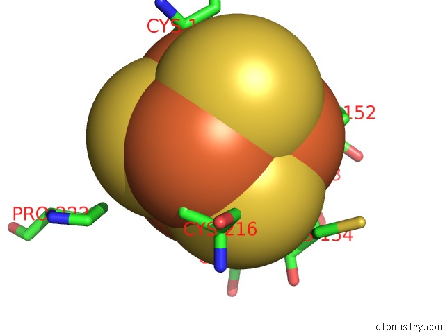
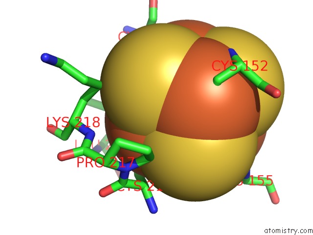
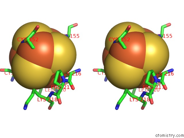
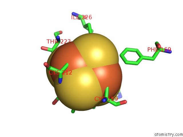
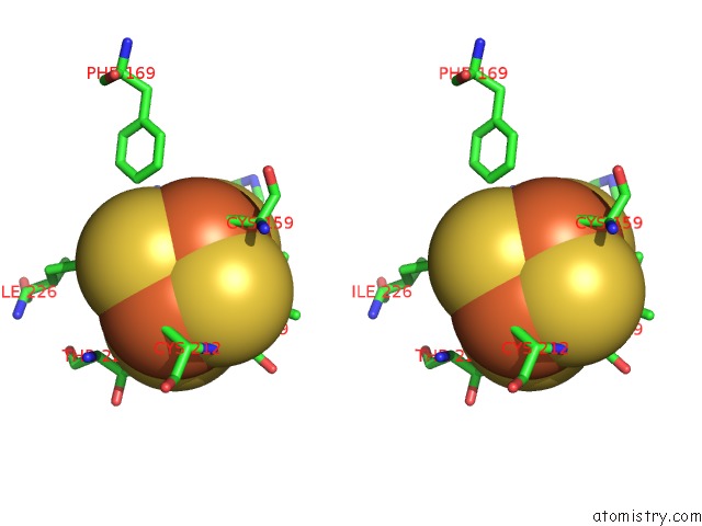
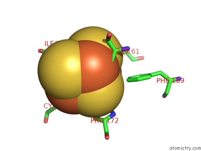
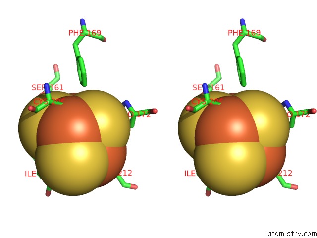
Iron binding site 1 out of 10 in 1nek
Go back to
Iron binding site 1 out
of 10 in the Complex II (Succinate Dehydrogenase) From E. Coli with Ubiquinone Bound
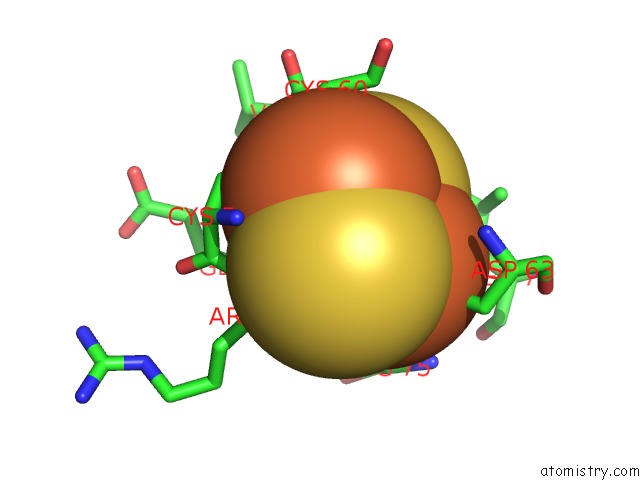
Mono view
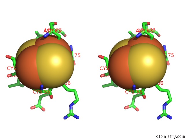
Stereo pair view
Mono view
Stereo pair view
A full contact list of Iron with other atoms in the Fe binding
site number 1 of Complex II (Succinate Dehydrogenase) From E. Coli with Ubiquinone Bound within 5.0Å range:
|
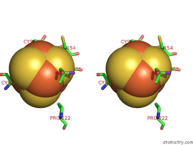
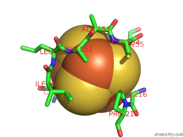
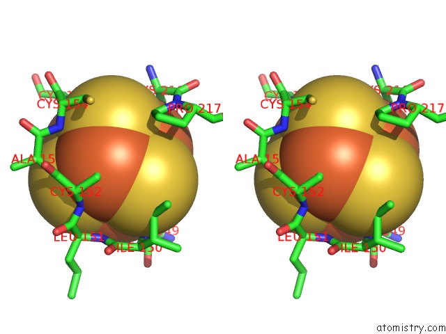
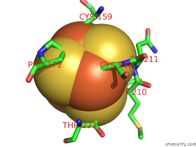
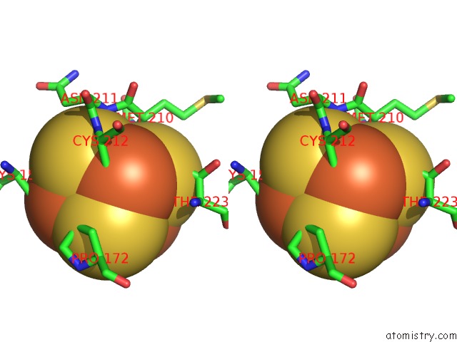
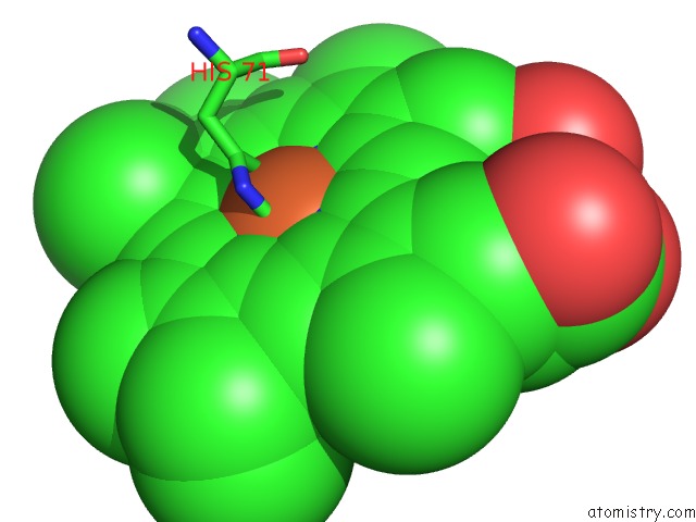
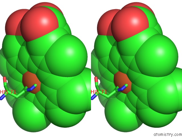
Iron binding site 2 out of 10 in 1nek
Go back to
Iron binding site 2 out
of 10 in the Complex II (Succinate Dehydrogenase) From E. Coli with Ubiquinone Bound
Mono view
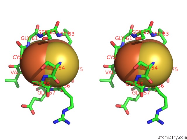
Stereo pair view
Mono view
Stereo pair view
A full contact list of Iron with other atoms in the Fe binding
site number 2 of Complex II (Succinate Dehydrogenase) From E. Coli with Ubiquinone Bound within 5.0Å range:
|
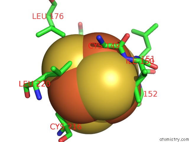
Iron binding site 3 out of 10 in 1nek
Go back to
Iron binding site 3 out
of 10 in the Complex II (Succinate Dehydrogenase) From E. Coli with Ubiquinone Bound
Mono view
Stereo pair view
Mono view
Stereo pair view
A full contact list of Iron with other atoms in the Fe binding
site number 3 of Complex II (Succinate Dehydrogenase) From E. Coli with Ubiquinone Bound within 5.0Å range:
|
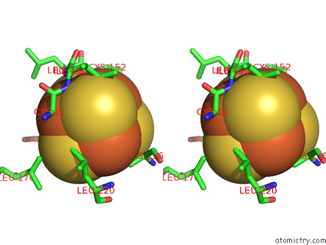
Iron binding site 4 out of 10 in 1nek
Go back to
Iron binding site 4 out
of 10 in the Complex II (Succinate Dehydrogenase) From E. Coli with Ubiquinone Bound
Mono view
Stereo pair view
Mono view
Stereo pair view
A full contact list of Iron with other atoms in the Fe binding
site number 4 of Complex II (Succinate Dehydrogenase) From E. Coli with Ubiquinone Bound within 5.0Å range:
|
Iron binding site 5 out of 10 in 1nek
Go back to
Iron binding site 5 out
of 10 in the Complex II (Succinate Dehydrogenase) From E. Coli with Ubiquinone Bound
Mono view
Stereo pair view
Mono view
Stereo pair view
A full contact list of Iron with other atoms in the Fe binding
site number 5 of Complex II (Succinate Dehydrogenase) From E. Coli with Ubiquinone Bound within 5.0Å range:
|
Iron binding site 6 out of 10 in 1nek
Go back to
Iron binding site 6 out
of 10 in the Complex II (Succinate Dehydrogenase) From E. Coli with Ubiquinone Bound
Mono view
Stereo pair view
Mono view
Stereo pair view
A full contact list of Iron with other atoms in the Fe binding
site number 6 of Complex II (Succinate Dehydrogenase) From E. Coli with Ubiquinone Bound within 5.0Å range:
|
Iron binding site 7 out of 10 in 1nek
Go back to
Iron binding site 7 out
of 10 in the Complex II (Succinate Dehydrogenase) From E. Coli with Ubiquinone Bound
Mono view
Stereo pair view
Mono view
Stereo pair view
A full contact list of Iron with other atoms in the Fe binding
site number 7 of Complex II (Succinate Dehydrogenase) From E. Coli with Ubiquinone Bound within 5.0Å range:
|
Iron binding site 8 out of 10 in 1nek
Go back to
Iron binding site 8 out
of 10 in the Complex II (Succinate Dehydrogenase) From E. Coli with Ubiquinone Bound
Mono view
Stereo pair view
Mono view
Stereo pair view
A full contact list of Iron with other atoms in the Fe binding
site number 8 of Complex II (Succinate Dehydrogenase) From E. Coli with Ubiquinone Bound within 5.0Å range:
|
Iron binding site 9 out of 10 in 1nek
Go back to
Iron binding site 9 out
of 10 in the Complex II (Succinate Dehydrogenase) From E. Coli with Ubiquinone Bound
Mono view
Stereo pair view
Mono view
Stereo pair view
A full contact list of Iron with other atoms in the Fe binding
site number 9 of Complex II (Succinate Dehydrogenase) From E. Coli with Ubiquinone Bound within 5.0Å range:
|
Iron binding site 10 out of 10 in 1nek
Go back to
Iron binding site 10 out
of 10 in the Complex II (Succinate Dehydrogenase) From E. Coli with Ubiquinone Bound
Mono view
Stereo pair view
Mono view
Stereo pair view
A full contact list of Iron with other atoms in the Fe binding
site number 10 of Complex II (Succinate Dehydrogenase) From E. Coli with Ubiquinone Bound within 5.0Å range:
|
Reference:
V.Yankovskaya,
R.Horsefield,
S.Tornroth,
C.Luna-Chavez,
H.Miyoshi,
C.Leger,
B.Byrne,
G.Cecchini,
S.Iwata.
Architecture of Succinate Dehydrogenase and Reactive Oxygen Species Generation. Science V. 299 700 2003.
ISSN: ISSN 0036-8075
PubMed: 12560550
DOI: 10.1126/SCIENCE.1079605
Page generated: Wed Jul 16 18:34:12 2025
ISSN: ISSN 0036-8075
PubMed: 12560550
DOI: 10.1126/SCIENCE.1079605
Last articles
Fe in 2YXOFe in 2YRS
Fe in 2YXC
Fe in 2YNM
Fe in 2YVJ
Fe in 2YP1
Fe in 2YU2
Fe in 2YU1
Fe in 2YQB
Fe in 2YOO