Iron »
PDB 1n5w-1nmi »
1nf4 »
Iron in PDB 1nf4: X-Ray Structure of the Desulfovibrio Desulfuricans Bacterioferritin: the Diiron Site in Different States (Reduced Structure)
Protein crystallography data
The structure of X-Ray Structure of the Desulfovibrio Desulfuricans Bacterioferritin: the Diiron Site in Different States (Reduced Structure), PDB code: 1nf4
was solved by
S.Macedo,
C.V.Romao,
E.Mitchell,
P.M.Matias,
M.Y.Liu,
A.V.Xavier,
J.Legall,
M.Teixeira,
P.Lindley,
M.A.Carrondo,
with X-Ray Crystallography technique. A brief refinement statistics is given in the table below:
| Resolution Low / High (Å) | 30.00 / 2.05 |
| Space group | P 21 3 1 |
| Cell size a, b, c (Å), α, β, γ (°) | 225.680, 225.680, 225.680, 90.00, 90.00, 90.00 |
| R / Rfree (%) | 23.1 / 27 |
Iron Binding Sites:
Pages:
>>> Page 1 <<< Page 2, Binding sites: 11 - 20; Page 3, Binding sites: 21 - 30; Page 4, Binding sites: 31 - 40; Page 5, Binding sites: 41 - 48;Binding sites:
The binding sites of Iron atom in the X-Ray Structure of the Desulfovibrio Desulfuricans Bacterioferritin: the Diiron Site in Different States (Reduced Structure) (pdb code 1nf4). This binding sites where shown within 5.0 Angstroms radius around Iron atom.In total 48 binding sites of Iron where determined in the X-Ray Structure of the Desulfovibrio Desulfuricans Bacterioferritin: the Diiron Site in Different States (Reduced Structure), PDB code: 1nf4:
Jump to Iron binding site number: 1; 2; 3; 4; 5; 6; 7; 8; 9; 10;
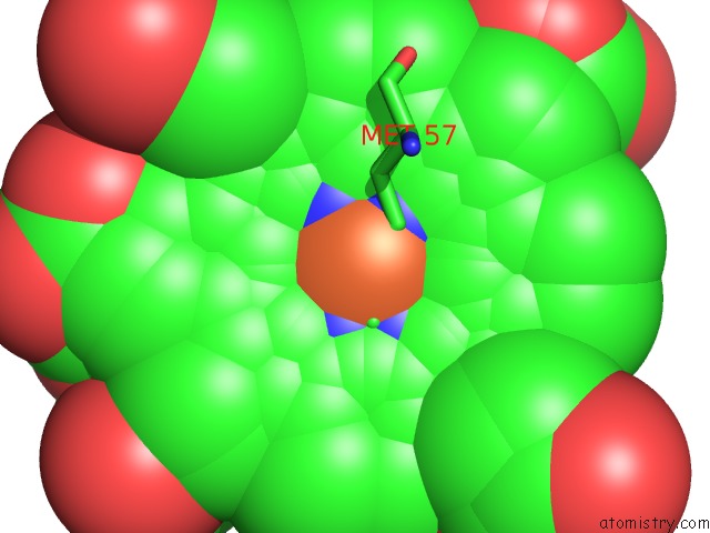
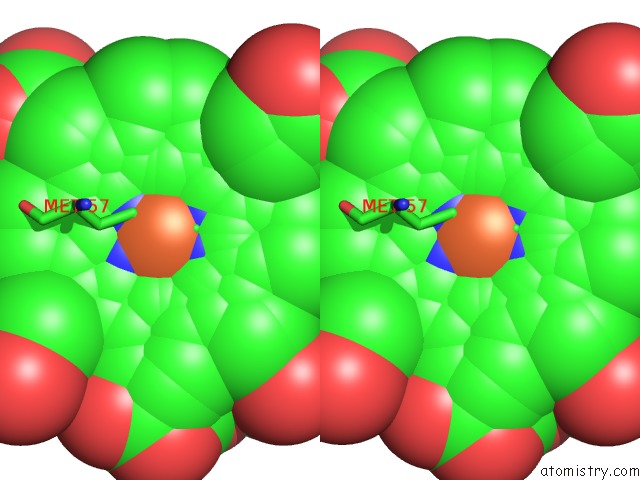
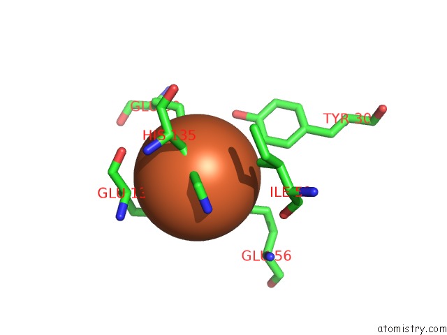
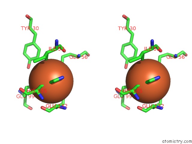
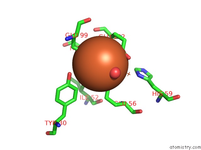
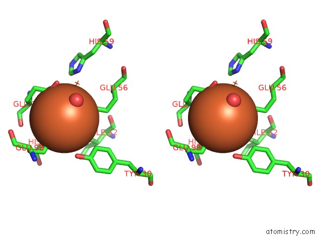
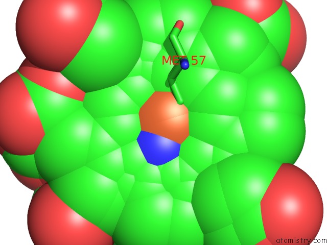
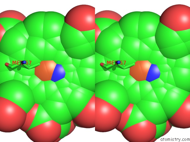
Iron binding site 1 out of 48 in 1nf4
Go back to
Iron binding site 1 out
of 48 in the X-Ray Structure of the Desulfovibrio Desulfuricans Bacterioferritin: the Diiron Site in Different States (Reduced Structure)
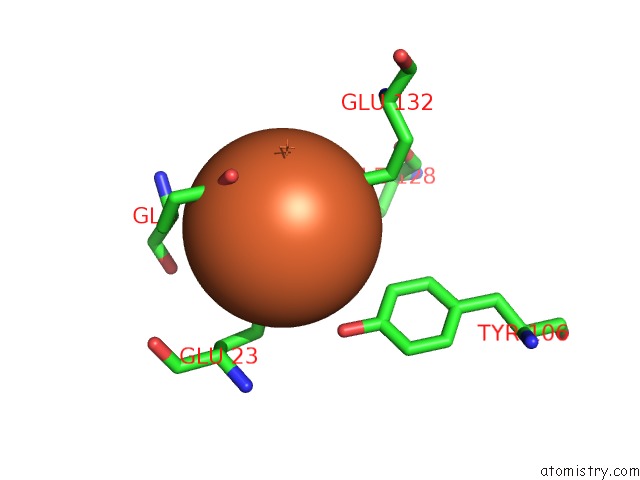
Mono view
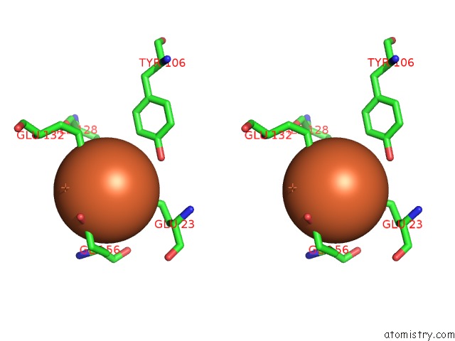
Stereo pair view
Mono view
Stereo pair view
A full contact list of Iron with other atoms in the Fe binding
site number 1 of X-Ray Structure of the Desulfovibrio Desulfuricans Bacterioferritin: the Diiron Site in Different States (Reduced Structure) within 5.0Å range:
|
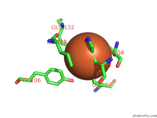
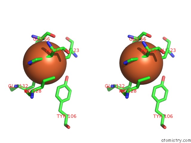
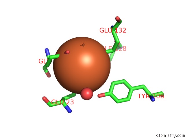
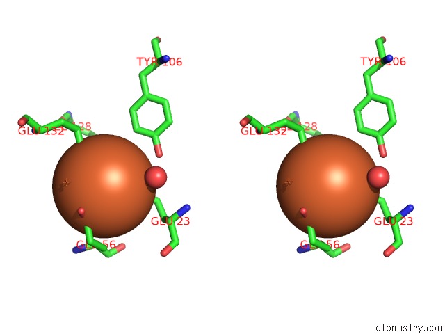
Iron binding site 2 out of 48 in 1nf4
Go back to
Iron binding site 2 out
of 48 in the X-Ray Structure of the Desulfovibrio Desulfuricans Bacterioferritin: the Diiron Site in Different States (Reduced Structure)
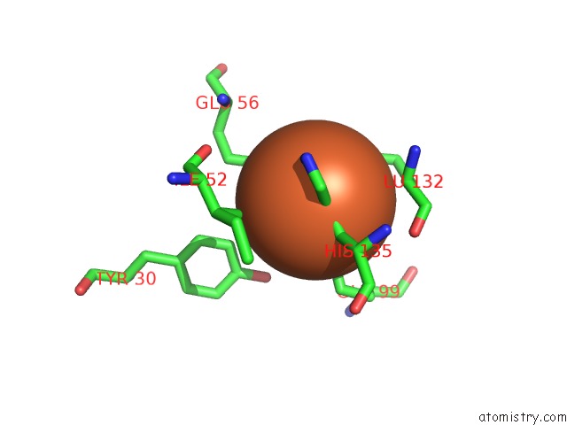
Mono view
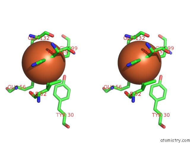
Stereo pair view
Mono view
Stereo pair view
A full contact list of Iron with other atoms in the Fe binding
site number 2 of X-Ray Structure of the Desulfovibrio Desulfuricans Bacterioferritin: the Diiron Site in Different States (Reduced Structure) within 5.0Å range:
|
Iron binding site 3 out of 48 in 1nf4
Go back to
Iron binding site 3 out
of 48 in the X-Ray Structure of the Desulfovibrio Desulfuricans Bacterioferritin: the Diiron Site in Different States (Reduced Structure)
Mono view

Stereo pair view
Mono view
Stereo pair view
A full contact list of Iron with other atoms in the Fe binding
site number 3 of X-Ray Structure of the Desulfovibrio Desulfuricans Bacterioferritin: the Diiron Site in Different States (Reduced Structure) within 5.0Å range:
|
Iron binding site 4 out of 48 in 1nf4
Go back to
Iron binding site 4 out
of 48 in the X-Ray Structure of the Desulfovibrio Desulfuricans Bacterioferritin: the Diiron Site in Different States (Reduced Structure)

Mono view
Stereo pair view
Mono view
Stereo pair view
A full contact list of Iron with other atoms in the Fe binding
site number 4 of X-Ray Structure of the Desulfovibrio Desulfuricans Bacterioferritin: the Diiron Site in Different States (Reduced Structure) within 5.0Å range:
|
Iron binding site 5 out of 48 in 1nf4
Go back to
Iron binding site 5 out
of 48 in the X-Ray Structure of the Desulfovibrio Desulfuricans Bacterioferritin: the Diiron Site in Different States (Reduced Structure)
Mono view
Stereo pair view
Mono view
Stereo pair view
A full contact list of Iron with other atoms in the Fe binding
site number 5 of X-Ray Structure of the Desulfovibrio Desulfuricans Bacterioferritin: the Diiron Site in Different States (Reduced Structure) within 5.0Å range:
|
Iron binding site 6 out of 48 in 1nf4
Go back to
Iron binding site 6 out
of 48 in the X-Ray Structure of the Desulfovibrio Desulfuricans Bacterioferritin: the Diiron Site in Different States (Reduced Structure)
Mono view
Stereo pair view
Mono view
Stereo pair view
A full contact list of Iron with other atoms in the Fe binding
site number 6 of X-Ray Structure of the Desulfovibrio Desulfuricans Bacterioferritin: the Diiron Site in Different States (Reduced Structure) within 5.0Å range:
|
Iron binding site 7 out of 48 in 1nf4
Go back to
Iron binding site 7 out
of 48 in the X-Ray Structure of the Desulfovibrio Desulfuricans Bacterioferritin: the Diiron Site in Different States (Reduced Structure)
Mono view
Stereo pair view
Mono view
Stereo pair view
A full contact list of Iron with other atoms in the Fe binding
site number 7 of X-Ray Structure of the Desulfovibrio Desulfuricans Bacterioferritin: the Diiron Site in Different States (Reduced Structure) within 5.0Å range:
|
Iron binding site 8 out of 48 in 1nf4
Go back to
Iron binding site 8 out
of 48 in the X-Ray Structure of the Desulfovibrio Desulfuricans Bacterioferritin: the Diiron Site in Different States (Reduced Structure)
Mono view
Stereo pair view
Mono view
Stereo pair view
A full contact list of Iron with other atoms in the Fe binding
site number 8 of X-Ray Structure of the Desulfovibrio Desulfuricans Bacterioferritin: the Diiron Site in Different States (Reduced Structure) within 5.0Å range:
|
Iron binding site 9 out of 48 in 1nf4
Go back to
Iron binding site 9 out
of 48 in the X-Ray Structure of the Desulfovibrio Desulfuricans Bacterioferritin: the Diiron Site in Different States (Reduced Structure)
Mono view

Stereo pair view
Mono view
Stereo pair view
A full contact list of Iron with other atoms in the Fe binding
site number 9 of X-Ray Structure of the Desulfovibrio Desulfuricans Bacterioferritin: the Diiron Site in Different States (Reduced Structure) within 5.0Å range:
|
Iron binding site 10 out of 48 in 1nf4
Go back to
Iron binding site 10 out
of 48 in the X-Ray Structure of the Desulfovibrio Desulfuricans Bacterioferritin: the Diiron Site in Different States (Reduced Structure)

Mono view
Stereo pair view
Mono view
Stereo pair view
A full contact list of Iron with other atoms in the Fe binding
site number 10 of X-Ray Structure of the Desulfovibrio Desulfuricans Bacterioferritin: the Diiron Site in Different States (Reduced Structure) within 5.0Å range:
|
Reference:
S.Macedo,
C.V.Romao,
E.Mitchell,
P.M.Matias,
M.Y.Liu,
A.V.Xavier,
J.Legall,
M.Teixeira,
P.Lindley,
M.A.Carrondo.
The Nature of the Di-Iron Site in the Bacterioferritin From Desulfovibrio Desulfuricans Nat.Struct.Biol. V. 10 285 2003.
ISSN: ISSN 1072-8368
PubMed: 12627224
DOI: 10.1038/NSB909
Page generated: Sat Aug 3 11:34:55 2024
ISSN: ISSN 1072-8368
PubMed: 12627224
DOI: 10.1038/NSB909
Last articles
F in 7Q2XF in 7PVK
F in 7Q2J
F in 7Q01
F in 7PZX
F in 7PZW
F in 7PZV
F in 7PZU
F in 7PZS
F in 7PY4