Iron »
PDB 1nml-1o1i »
1nnf »
Iron in PDB 1nnf: Crystal Structure Analysis of Haemophlius Influenzae Ferric- Ion Binding Protein H9Q Mutant Form
Protein crystallography data
The structure of Crystal Structure Analysis of Haemophlius Influenzae Ferric- Ion Binding Protein H9Q Mutant Form, PDB code: 1nnf
was solved by
S.R.Shouldice,
D.R.Dougan,
R.J.Skene,
L.W.Tari,
D.E.Mcree,
R.-H.Yu,
A.B.Schryvers,
with X-Ray Crystallography technique. A brief refinement statistics is given in the table below:
| Resolution Low / High (Å) | 105.41 / 1.10 |
| Space group | P 21 21 2 |
| Cell size a, b, c (Å), α, β, γ (°) | 105.101, 75.344, 33.503, 90.00, 90.00, 90.00 |
| R / Rfree (%) | 15.8 / 18 |
Iron Binding Sites:
The binding sites of Iron atom in the Crystal Structure Analysis of Haemophlius Influenzae Ferric- Ion Binding Protein H9Q Mutant Form
(pdb code 1nnf). This binding sites where shown within
5.0 Angstroms radius around Iron atom.
In total only one binding site of Iron was determined in the Crystal Structure Analysis of Haemophlius Influenzae Ferric- Ion Binding Protein H9Q Mutant Form, PDB code: 1nnf:
In total only one binding site of Iron was determined in the Crystal Structure Analysis of Haemophlius Influenzae Ferric- Ion Binding Protein H9Q Mutant Form, PDB code: 1nnf:
Iron binding site 1 out of 1 in 1nnf
Go back to
Iron binding site 1 out
of 1 in the Crystal Structure Analysis of Haemophlius Influenzae Ferric- Ion Binding Protein H9Q Mutant Form
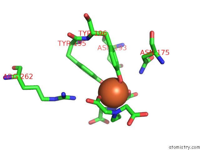
Mono view
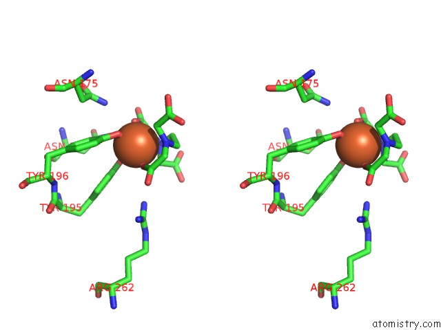
Stereo pair view
Mono view
Stereo pair view
A full contact list of Iron with other atoms in the Fe binding
site number 1 of Crystal Structure Analysis of Haemophlius Influenzae Ferric- Ion Binding Protein H9Q Mutant Form within 5.0Å range:
|
Reference:
S.R.Shouldice,
D.R.Dougan,
R.J.Skene,
L.W.Tari,
D.E.Mcree,
R.-H.Yu,
A.B.Schryvers.
High Resolution Structure of An Alternate Form of the Ferric Ion Binding Protein From Haemophilus Influenzae J.Biol.Chem. V. 278 11513 2003.
ISSN: ISSN 0021-9258
PubMed: 12533539
DOI: 10.1074/JBC.M211780200
Page generated: Sat Aug 3 11:57:17 2024
ISSN: ISSN 0021-9258
PubMed: 12533539
DOI: 10.1074/JBC.M211780200
Last articles
Zn in 9J0NZn in 9J0O
Zn in 9J0P
Zn in 9FJX
Zn in 9EKB
Zn in 9C0F
Zn in 9CAH
Zn in 9CH0
Zn in 9CH3
Zn in 9CH1