Iron »
PDB 1nml-1o1i »
1np1 »
Iron in PDB 1np1: Crystal Structure of the Complex of Nitrophorin 1 From Rhodnius Prolixus with Histamine
Protein crystallography data
The structure of Crystal Structure of the Complex of Nitrophorin 1 From Rhodnius Prolixus with Histamine, PDB code: 1np1
was solved by
A.Weichsel,
W.R.Montfort,
with X-Ray Crystallography technique. A brief refinement statistics is given in the table below:
| Resolution Low / High (Å) | 15.00 / 2.00 |
| Space group | P 1 21 1 |
| Cell size a, b, c (Å), α, β, γ (°) | 39.550, 74.570, 66.560, 90.00, 99.94, 90.00 |
| R / Rfree (%) | 18.1 / 27.8 |
Iron Binding Sites:
The binding sites of Iron atom in the Crystal Structure of the Complex of Nitrophorin 1 From Rhodnius Prolixus with Histamine
(pdb code 1np1). This binding sites where shown within
5.0 Angstroms radius around Iron atom.
In total 2 binding sites of Iron where determined in the Crystal Structure of the Complex of Nitrophorin 1 From Rhodnius Prolixus with Histamine, PDB code: 1np1:
Jump to Iron binding site number: 1; 2;
In total 2 binding sites of Iron where determined in the Crystal Structure of the Complex of Nitrophorin 1 From Rhodnius Prolixus with Histamine, PDB code: 1np1:
Jump to Iron binding site number: 1; 2;
Iron binding site 1 out of 2 in 1np1
Go back to
Iron binding site 1 out
of 2 in the Crystal Structure of the Complex of Nitrophorin 1 From Rhodnius Prolixus with Histamine
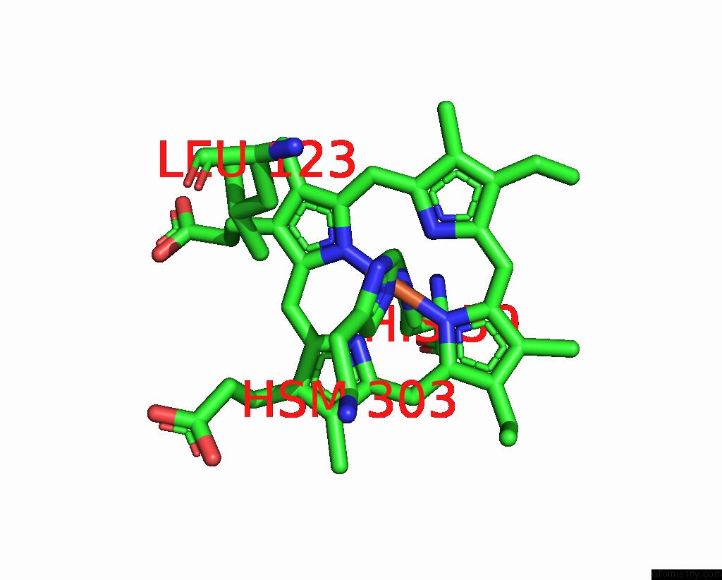
Mono view
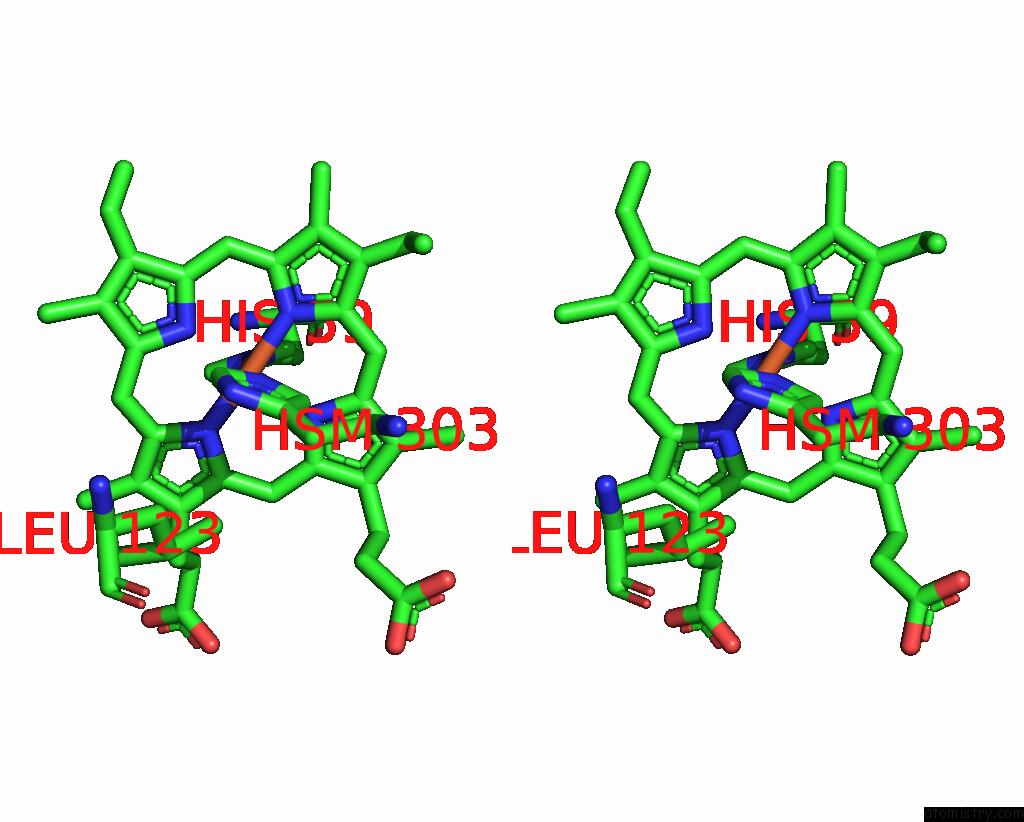
Stereo pair view
Mono view
Stereo pair view
A full contact list of Iron with other atoms in the Fe binding
site number 1 of Crystal Structure of the Complex of Nitrophorin 1 From Rhodnius Prolixus with Histamine within 5.0Å range:
|
Iron binding site 2 out of 2 in 1np1
Go back to
Iron binding site 2 out
of 2 in the Crystal Structure of the Complex of Nitrophorin 1 From Rhodnius Prolixus with Histamine
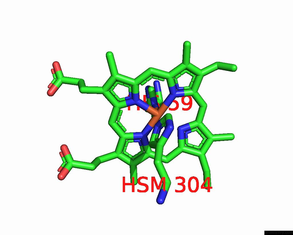
Mono view
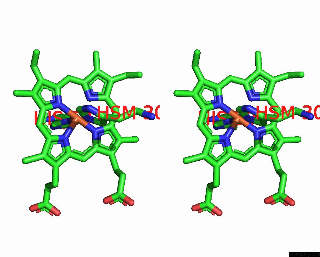
Stereo pair view
Mono view
Stereo pair view
A full contact list of Iron with other atoms in the Fe binding
site number 2 of Crystal Structure of the Complex of Nitrophorin 1 From Rhodnius Prolixus with Histamine within 5.0Å range:
|
Reference:
A.Weichsel,
J.F.Andersen,
D.E.Champagne,
F.A.Walker,
W.R.Montfort.
Crystal Structures of A Nitric Oxide Transport Protein From A Blood-Sucking Insect. Nat.Struct.Biol. V. 5 304 1998.
ISSN: ISSN 1072-8368
PubMed: 9546222
DOI: 10.1038/NSB0498-304
Page generated: Sat Aug 3 11:58:16 2024
ISSN: ISSN 1072-8368
PubMed: 9546222
DOI: 10.1038/NSB0498-304
Last articles
Zn in 9J0NZn in 9J0O
Zn in 9J0P
Zn in 9FJX
Zn in 9EKB
Zn in 9C0F
Zn in 9CAH
Zn in 9CH0
Zn in 9CH3
Zn in 9CH1