Iron »
PDB 1nml-1o1i »
1ntk »
Iron in PDB 1ntk: Crystal Structure of Mitochondrial Cytochrome BC1 in Complex with Antimycin A1
Enzymatic activity of Crystal Structure of Mitochondrial Cytochrome BC1 in Complex with Antimycin A1
All present enzymatic activity of Crystal Structure of Mitochondrial Cytochrome BC1 in Complex with Antimycin A1:
1.10.2.2;
1.10.2.2;
Protein crystallography data
The structure of Crystal Structure of Mitochondrial Cytochrome BC1 in Complex with Antimycin A1, PDB code: 1ntk
was solved by
X.Gao,
X.Wen,
L.Esser,
B.Quinn,
L.Yu,
C.-A.Yu,
D.Xia,
with X-Ray Crystallography technique. A brief refinement statistics is given in the table below:
| Resolution Low / High (Å) | 40.00 / 2.60 |
| Space group | I 41 2 2 |
| Cell size a, b, c (Å), α, β, γ (°) | 153.785, 153.785, 592.498, 90.00, 90.00, 90.00 |
| R / Rfree (%) | 23.3 / 27 |
Iron Binding Sites:
The binding sites of Iron atom in the Crystal Structure of Mitochondrial Cytochrome BC1 in Complex with Antimycin A1
(pdb code 1ntk). This binding sites where shown within
5.0 Angstroms radius around Iron atom.
In total 5 binding sites of Iron where determined in the Crystal Structure of Mitochondrial Cytochrome BC1 in Complex with Antimycin A1, PDB code: 1ntk:
Jump to Iron binding site number: 1; 2; 3; 4; 5;
In total 5 binding sites of Iron where determined in the Crystal Structure of Mitochondrial Cytochrome BC1 in Complex with Antimycin A1, PDB code: 1ntk:
Jump to Iron binding site number: 1; 2; 3; 4; 5;
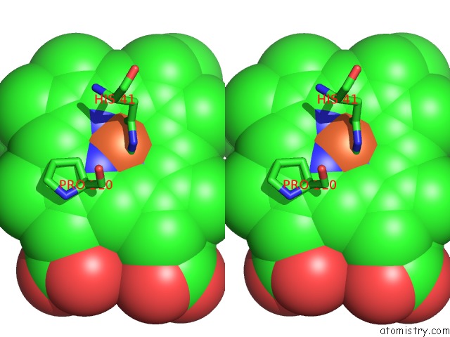
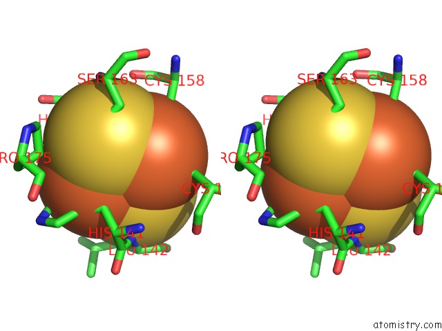
Iron binding site 1 out of 5 in 1ntk
Go back to
Iron binding site 1 out
of 5 in the Crystal Structure of Mitochondrial Cytochrome BC1 in Complex with Antimycin A1

Mono view
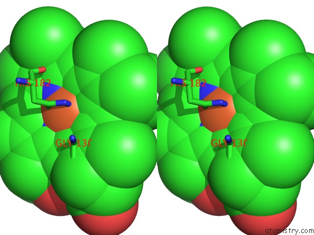
Stereo pair view
Mono view
Stereo pair view
A full contact list of Iron with other atoms in the Fe binding
site number 1 of Crystal Structure of Mitochondrial Cytochrome BC1 in Complex with Antimycin A1 within 5.0Å range:
|
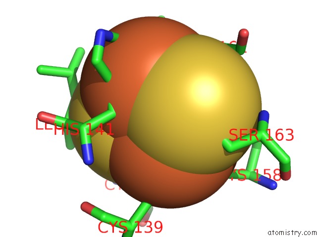
Iron binding site 2 out of 5 in 1ntk
Go back to
Iron binding site 2 out
of 5 in the Crystal Structure of Mitochondrial Cytochrome BC1 in Complex with Antimycin A1
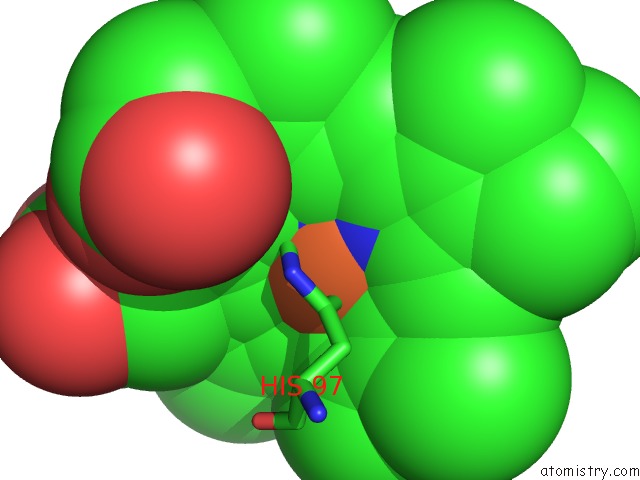
Mono view
Stereo pair view
Mono view
Stereo pair view
A full contact list of Iron with other atoms in the Fe binding
site number 2 of Crystal Structure of Mitochondrial Cytochrome BC1 in Complex with Antimycin A1 within 5.0Å range:
|
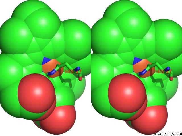
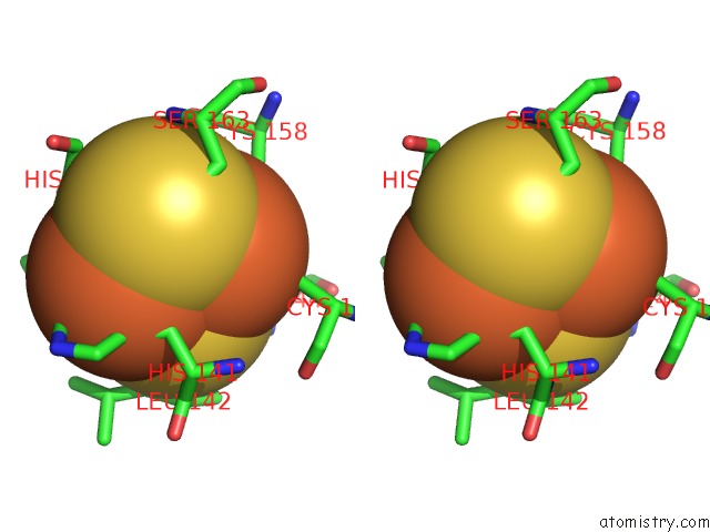
Iron binding site 3 out of 5 in 1ntk
Go back to
Iron binding site 3 out
of 5 in the Crystal Structure of Mitochondrial Cytochrome BC1 in Complex with Antimycin A1
Mono view
Stereo pair view
Mono view
Stereo pair view
A full contact list of Iron with other atoms in the Fe binding
site number 3 of Crystal Structure of Mitochondrial Cytochrome BC1 in Complex with Antimycin A1 within 5.0Å range:
|
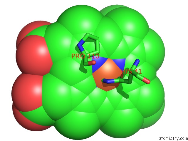
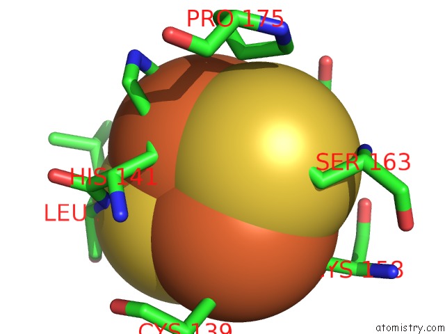
Iron binding site 4 out of 5 in 1ntk
Go back to
Iron binding site 4 out
of 5 in the Crystal Structure of Mitochondrial Cytochrome BC1 in Complex with Antimycin A1
Mono view
Stereo pair view
Mono view
Stereo pair view
A full contact list of Iron with other atoms in the Fe binding
site number 4 of Crystal Structure of Mitochondrial Cytochrome BC1 in Complex with Antimycin A1 within 5.0Å range:
|
Iron binding site 5 out of 5 in 1ntk
Go back to
Iron binding site 5 out
of 5 in the Crystal Structure of Mitochondrial Cytochrome BC1 in Complex with Antimycin A1
Mono view
Stereo pair view
Mono view
Stereo pair view
A full contact list of Iron with other atoms in the Fe binding
site number 5 of Crystal Structure of Mitochondrial Cytochrome BC1 in Complex with Antimycin A1 within 5.0Å range:
|
Reference:
X.Gao,
X.Wen,
L.Esser,
B.Quinn,
L.Yu,
C.-A.Yu,
D.Xia.
Structural Basis For the Quinone Reduction in the Bc(1) Complex: A Comparative Analysis of Crystal Structures of Mitochondrial Cytochrome Bc(1) with Bound Substrate and Inhibitors at the Q(I) Site Biochemistry V. 42 9067 2003.
ISSN: ISSN 0006-2960
PubMed: 12885240
DOI: 10.1021/BI0341814
Page generated: Sat Aug 3 12:00:34 2024
ISSN: ISSN 0006-2960
PubMed: 12885240
DOI: 10.1021/BI0341814
Last articles
Cl in 3OJNCl in 3OJK
Cl in 3OJJ
Cl in 3OIF
Cl in 3OJ8
Cl in 3OIR
Cl in 3OJ6
Cl in 3OIY
Cl in 3OIN
Cl in 3OIO