Iron »
PDB 1nml-1o1i »
1nwn »
Iron in PDB 1nwn: Crystals of Co-Hbi in Which the Structure Was Converted to Its Unligated State, and Then Converted Back to Its Original Co-Ligated State.
Protein crystallography data
The structure of Crystals of Co-Hbi in Which the Structure Was Converted to Its Unligated State, and Then Converted Back to Its Original Co-Ligated State., PDB code: 1nwn
was solved by
J.E.Knapp,
W.E.Royer Jr.,
with X-Ray Crystallography technique. A brief refinement statistics is given in the table below:
| Resolution Low / High (Å) | 37.79 / 2.80 |
| Space group | C 1 2 1 |
| Cell size a, b, c (Å), α, β, γ (°) | 92.620, 43.510, 83.110, 90.00, 121.88, 90.00 |
| R / Rfree (%) | 22.2 / 24.8 |
Iron Binding Sites:
The binding sites of Iron atom in the Crystals of Co-Hbi in Which the Structure Was Converted to Its Unligated State, and Then Converted Back to Its Original Co-Ligated State.
(pdb code 1nwn). This binding sites where shown within
5.0 Angstroms radius around Iron atom.
In total 2 binding sites of Iron where determined in the Crystals of Co-Hbi in Which the Structure Was Converted to Its Unligated State, and Then Converted Back to Its Original Co-Ligated State., PDB code: 1nwn:
Jump to Iron binding site number: 1; 2;
In total 2 binding sites of Iron where determined in the Crystals of Co-Hbi in Which the Structure Was Converted to Its Unligated State, and Then Converted Back to Its Original Co-Ligated State., PDB code: 1nwn:
Jump to Iron binding site number: 1; 2;
Iron binding site 1 out of 2 in 1nwn
Go back to
Iron binding site 1 out
of 2 in the Crystals of Co-Hbi in Which the Structure Was Converted to Its Unligated State, and Then Converted Back to Its Original Co-Ligated State.
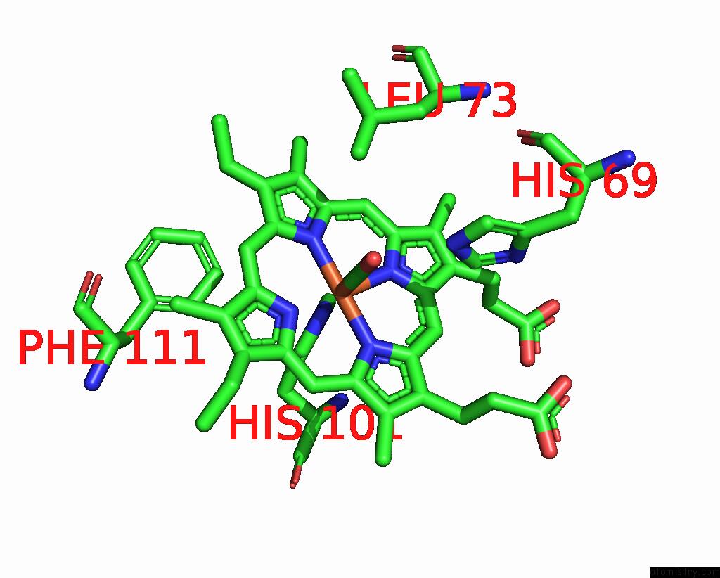
Mono view
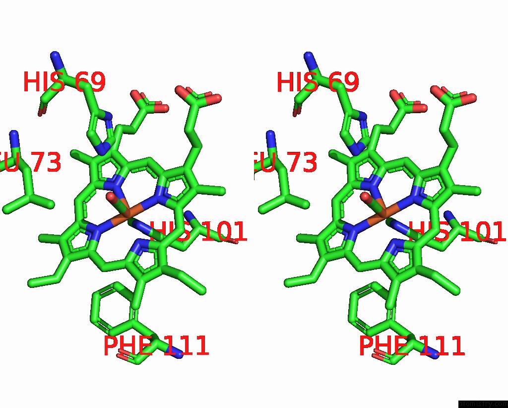
Stereo pair view
Mono view
Stereo pair view
A full contact list of Iron with other atoms in the Fe binding
site number 1 of Crystals of Co-Hbi in Which the Structure Was Converted to Its Unligated State, and Then Converted Back to Its Original Co-Ligated State. within 5.0Å range:
|
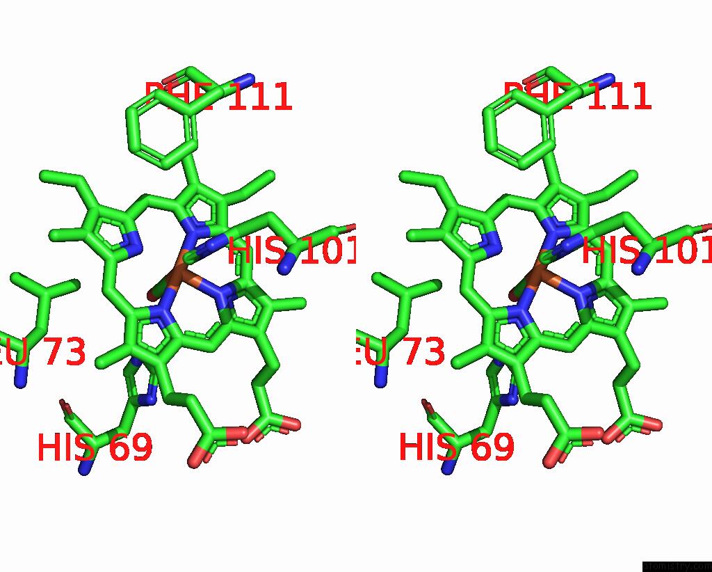
Iron binding site 2 out of 2 in 1nwn
Go back to
Iron binding site 2 out
of 2 in the Crystals of Co-Hbi in Which the Structure Was Converted to Its Unligated State, and Then Converted Back to Its Original Co-Ligated State.

Mono view
Stereo pair view
Mono view
Stereo pair view
A full contact list of Iron with other atoms in the Fe binding
site number 2 of Crystals of Co-Hbi in Which the Structure Was Converted to Its Unligated State, and Then Converted Back to Its Original Co-Ligated State. within 5.0Å range:
|
Reference:
J.E.Knapp,
W.E.Royer Jr..
Ligand-Linked Structural Transitions in Crystals of A Cooperative Dimeric Hemoglobin. Biochemistry V. 42 4640 2003.
ISSN: ISSN 0006-2960
PubMed: 12705827
DOI: 10.1021/BI027136P
Page generated: Wed Jul 16 18:55:08 2025
ISSN: ISSN 0006-2960
PubMed: 12705827
DOI: 10.1021/BI027136P
Last articles
Fe in 2YXOFe in 2YRS
Fe in 2YXC
Fe in 2YNM
Fe in 2YVJ
Fe in 2YP1
Fe in 2YU2
Fe in 2YU1
Fe in 2YQB
Fe in 2YOO