Iron »
PDB 1nml-1o1i »
1nz3 »
Iron in PDB 1nz3: K45E-K63E Variant of Horse Heart Myoglobin
Protein crystallography data
The structure of K45E-K63E Variant of Horse Heart Myoglobin, PDB code: 1nz3
was solved by
C.L.Hunter,
R.Maurus,
M.R.Mauk,
H.Lee,
E.L.Raven,
H.Tong,
N.Nguyen,
S.Smith,
G.D.Brayer,
A.G.Mauk,
with X-Ray Crystallography technique. A brief refinement statistics is given in the table below:
| Resolution Low / High (Å) | 10.00 / 1.60 |
| Space group | P 21 21 21 |
| Cell size a, b, c (Å), α, β, γ (°) | 29.000, 35.800, 125.800, 90.00, 90.00, 90.00 |
| R / Rfree (%) | n/a / n/a |
Iron Binding Sites:
The binding sites of Iron atom in the K45E-K63E Variant of Horse Heart Myoglobin
(pdb code 1nz3). This binding sites where shown within
5.0 Angstroms radius around Iron atom.
In total only one binding site of Iron was determined in the K45E-K63E Variant of Horse Heart Myoglobin, PDB code: 1nz3:
In total only one binding site of Iron was determined in the K45E-K63E Variant of Horse Heart Myoglobin, PDB code: 1nz3:
Iron binding site 1 out of 1 in 1nz3
Go back to
Iron binding site 1 out
of 1 in the K45E-K63E Variant of Horse Heart Myoglobin
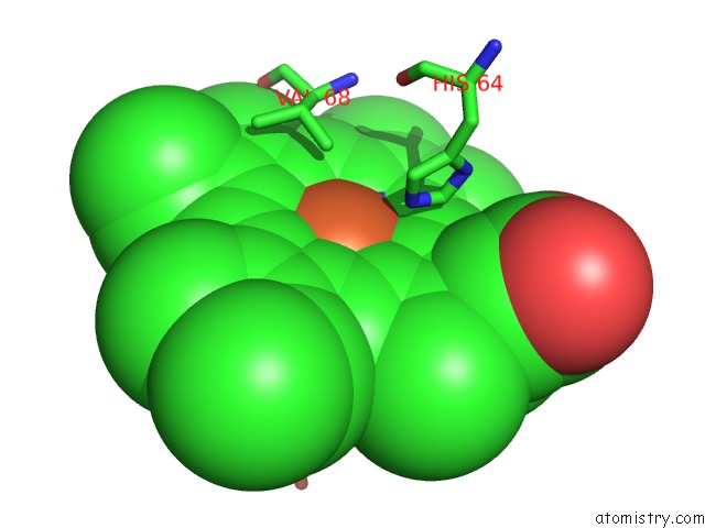
Mono view
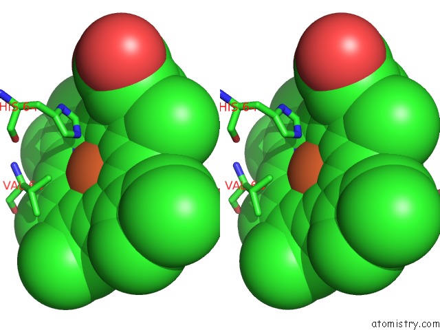
Stereo pair view
Mono view
Stereo pair view
A full contact list of Iron with other atoms in the Fe binding
site number 1 of K45E-K63E Variant of Horse Heart Myoglobin within 5.0Å range:
|
Reference:
C.L.Hunter,
R.Maurus,
M.R.Mauk,
H.Lee,
E.L.Raven,
H.Tong,
N.Nguyen,
S.Smith,
G.D.Brayer,
A.G.Mauk.
Introduction and Characterization of A Functionally Linked Metal Ion Binding Site at the Exposed Heme Edge of Myoglobin Proc.Natl.Acad.Sci.Usa V. 100 3647 2003.
ISSN: ISSN 0027-8424
PubMed: 12644706
DOI: 10.1073/PNAS.0636702100
Page generated: Sat Aug 3 12:05:37 2024
ISSN: ISSN 0027-8424
PubMed: 12644706
DOI: 10.1073/PNAS.0636702100
Last articles
Zn in 9J0NZn in 9J0O
Zn in 9J0P
Zn in 9FJX
Zn in 9EKB
Zn in 9C0F
Zn in 9CAH
Zn in 9CH0
Zn in 9CH3
Zn in 9CH1