Iron »
PDB 1o1j-1off »
1occ »
Iron in PDB 1occ: Structure of Bovine Heart Cytochrome C Oxidase at the Fully Oxidized State
Enzymatic activity of Structure of Bovine Heart Cytochrome C Oxidase at the Fully Oxidized State
All present enzymatic activity of Structure of Bovine Heart Cytochrome C Oxidase at the Fully Oxidized State:
1.9.3.1;
1.9.3.1;
Protein crystallography data
The structure of Structure of Bovine Heart Cytochrome C Oxidase at the Fully Oxidized State, PDB code: 1occ
was solved by
T.Tsukihara,
H.Aoyama,
E.Yamashita,
T.Tomizaki,
H.Yamaguchi,
K.Shinzawa-Itoh,
R.Nakashima,
R.Yaono,
S.Yoshikawa,
with X-Ray Crystallography technique. A brief refinement statistics is given in the table below:
| Resolution Low / High (Å) | 10.00 / 2.80 |
| Space group | P 21 21 21 |
| Cell size a, b, c (Å), α, β, γ (°) | 189.100, 210.500, 178.600, 90.00, 90.00, 90.00 |
| R / Rfree (%) | 20.1 / 25.2 |
Other elements in 1occ:
The structure of Structure of Bovine Heart Cytochrome C Oxidase at the Fully Oxidized State also contains other interesting chemical elements:
| Magnesium | (Mg) | 2 atoms |
| Copper | (Cu) | 6 atoms |
| Zinc | (Zn) | 2 atoms |
Iron Binding Sites:
The binding sites of Iron atom in the Structure of Bovine Heart Cytochrome C Oxidase at the Fully Oxidized State
(pdb code 1occ). This binding sites where shown within
5.0 Angstroms radius around Iron atom.
In total 4 binding sites of Iron where determined in the Structure of Bovine Heart Cytochrome C Oxidase at the Fully Oxidized State, PDB code: 1occ:
Jump to Iron binding site number: 1; 2; 3; 4;
In total 4 binding sites of Iron where determined in the Structure of Bovine Heart Cytochrome C Oxidase at the Fully Oxidized State, PDB code: 1occ:
Jump to Iron binding site number: 1; 2; 3; 4;
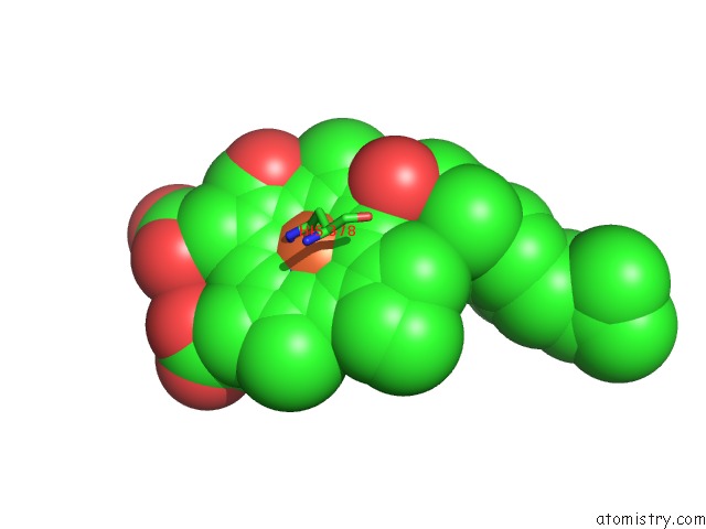
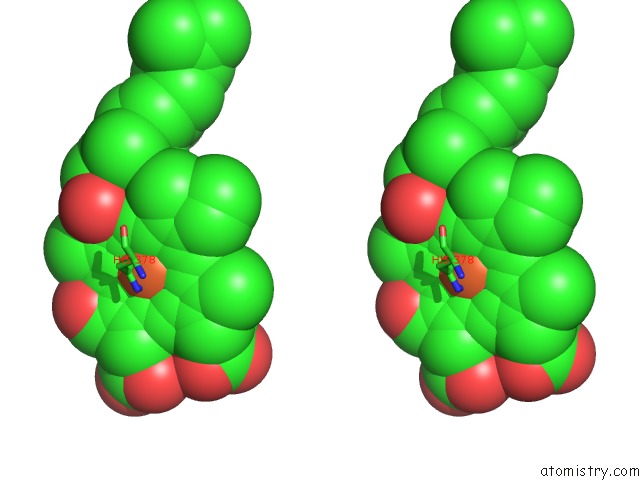
Iron binding site 1 out of 4 in 1occ
Go back to
Iron binding site 1 out
of 4 in the Structure of Bovine Heart Cytochrome C Oxidase at the Fully Oxidized State
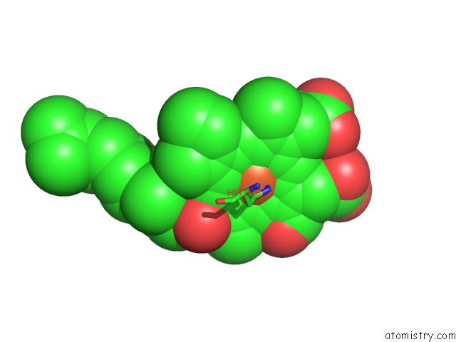
Mono view
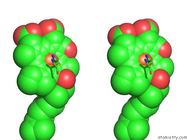
Stereo pair view
Mono view
Stereo pair view
A full contact list of Iron with other atoms in the Fe binding
site number 1 of Structure of Bovine Heart Cytochrome C Oxidase at the Fully Oxidized State within 5.0Å range:
|
Iron binding site 2 out of 4 in 1occ
Go back to
Iron binding site 2 out
of 4 in the Structure of Bovine Heart Cytochrome C Oxidase at the Fully Oxidized State
Mono view
Stereo pair view
Mono view
Stereo pair view
A full contact list of Iron with other atoms in the Fe binding
site number 2 of Structure of Bovine Heart Cytochrome C Oxidase at the Fully Oxidized State within 5.0Å range:
|
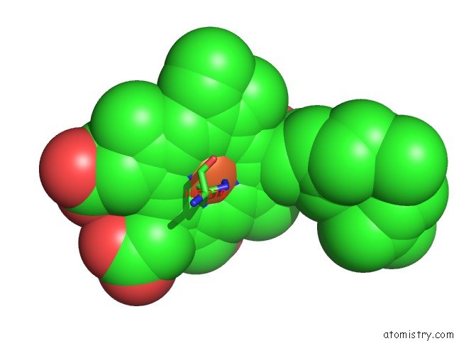
Iron binding site 3 out of 4 in 1occ
Go back to
Iron binding site 3 out
of 4 in the Structure of Bovine Heart Cytochrome C Oxidase at the Fully Oxidized State
Mono view
Stereo pair view
Mono view
Stereo pair view
A full contact list of Iron with other atoms in the Fe binding
site number 3 of Structure of Bovine Heart Cytochrome C Oxidase at the Fully Oxidized State within 5.0Å range:
|
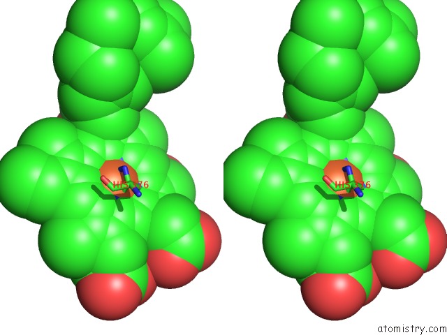
Iron binding site 4 out of 4 in 1occ
Go back to
Iron binding site 4 out
of 4 in the Structure of Bovine Heart Cytochrome C Oxidase at the Fully Oxidized State
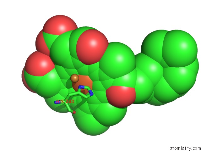
Mono view
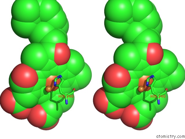
Stereo pair view
Mono view
Stereo pair view
A full contact list of Iron with other atoms in the Fe binding
site number 4 of Structure of Bovine Heart Cytochrome C Oxidase at the Fully Oxidized State within 5.0Å range:
|
Reference:
T.Tsukihara,
H.Aoyama,
E.Yamashita,
T.Tomizaki,
H.Yamaguchi,
K.Shinzawa-Itoh,
R.Nakashima,
R.Yaono,
S.Yoshikawa.
The Whole Structure of the 13-Subunit Oxidized Cytochrome C Oxidase at 2.8 A. Science V. 272 1136 1996.
ISSN: ISSN 0036-8075
PubMed: 8638158
Page generated: Wed Jul 16 19:06:22 2025
ISSN: ISSN 0036-8075
PubMed: 8638158
Last articles
Fe in 2YXOFe in 2YRS
Fe in 2YXC
Fe in 2YNM
Fe in 2YVJ
Fe in 2YP1
Fe in 2YU2
Fe in 2YU1
Fe in 2YQB
Fe in 2YOO