Iron »
PDB 1o1j-1off »
1odo »
Iron in PDB 1odo: 1.85 A Structure of CYP154A1 From Streptomyces Coelicolor A3(2)
Protein crystallography data
The structure of 1.85 A Structure of CYP154A1 From Streptomyces Coelicolor A3(2), PDB code: 1odo
was solved by
L.M.Podust,
Y.Kim,
M.Arase,
H.Bach,
D.H.Sherman,
D.C.Lamb,
S.L.Kelly,
M.R.Waterman,
with X-Ray Crystallography technique. A brief refinement statistics is given in the table below:
| Resolution Low / High (Å) | 33.21 / 1.85 |
| Space group | P 21 21 21 |
| Cell size a, b, c (Å), α, β, γ (°) | 47.510, 103.708, 104.310, 90.00, 90.00, 90.00 |
| R / Rfree (%) | 17.7 / 20.8 |
Iron Binding Sites:
The binding sites of Iron atom in the 1.85 A Structure of CYP154A1 From Streptomyces Coelicolor A3(2)
(pdb code 1odo). This binding sites where shown within
5.0 Angstroms radius around Iron atom.
In total only one binding site of Iron was determined in the 1.85 A Structure of CYP154A1 From Streptomyces Coelicolor A3(2), PDB code: 1odo:
In total only one binding site of Iron was determined in the 1.85 A Structure of CYP154A1 From Streptomyces Coelicolor A3(2), PDB code: 1odo:
Iron binding site 1 out of 1 in 1odo
Go back to
Iron binding site 1 out
of 1 in the 1.85 A Structure of CYP154A1 From Streptomyces Coelicolor A3(2)
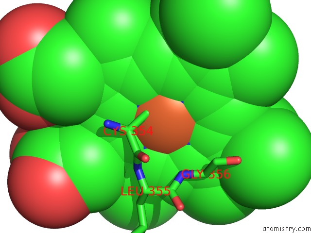
Mono view
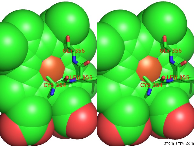
Stereo pair view
Mono view
Stereo pair view
A full contact list of Iron with other atoms in the Fe binding
site number 1 of 1.85 A Structure of CYP154A1 From Streptomyces Coelicolor A3(2) within 5.0Å range:
|
Reference:
L.M.Podust,
H.Bach,
Y.Kim,
D.C.Lamb,
M.Arase,
D.H.Sherman,
S.L.Kelly,
M.R.Waterman.
Comparison of the 1.85 A Structure of CYP154A1 From Streptomyces Coelicolor A3(2) with the Closely Related CYP154C1 and Cyps From Antibiotic Biosynthetic Pathways. Protein Sci. V. 13 255 2004.
ISSN: ISSN 0961-8368
PubMed: 14691240
DOI: 10.1110/PS.03384804
Page generated: Sat Aug 3 12:19:44 2024
ISSN: ISSN 0961-8368
PubMed: 14691240
DOI: 10.1110/PS.03384804
Last articles
Zn in 9MJ5Zn in 9HNW
Zn in 9G0L
Zn in 9FNE
Zn in 9DZN
Zn in 9E0I
Zn in 9D32
Zn in 9DAK
Zn in 8ZXC
Zn in 8ZUF