Atomistry » Iron » PDB 1ofj-1ozl » 1ogy
Atomistry »
  Iron »
    PDB 1ofj-1ozl »
      1ogy »

Iron in PDB 1ogy: Crystal Structure of the Heterodimeric Nitrate Reductase From Rhodobacter Sphaeroides

Enzymatic activity of Crystal Structure of the Heterodimeric Nitrate Reductase From Rhodobacter Sphaeroides

All present enzymatic activity of Crystal Structure of the Heterodimeric Nitrate Reductase From Rhodobacter Sphaeroides:
1.7.99.4;

Protein crystallography data

The structure of Crystal Structure of the Heterodimeric Nitrate Reductase From Rhodobacter Sphaeroides, PDB code: 1ogy was solved by P.Arnoux, M.Sabaty, J.Alric, B.Frangioni, B.Guigliarelli, J.-M.Adriano, D.Pignol, with X-Ray Crystallography technique. A brief refinement statistics is given in the table below:

Resolution Low / High (Å) 30.00 / 3.20
Space group P 1 21 1
Cell size a, b, c (Å), α, β, γ (°) 123.000, 225.200, 154.600, 90.00, 92.10, 90.00
R / Rfree (%) 25 / 26.9

Other elements in 1ogy:

The structure of Crystal Structure of the Heterodimeric Nitrate Reductase From Rhodobacter Sphaeroides also contains other interesting chemical elements:

Molybdenum (Mo) 8 atoms

Iron Binding Sites:

Pages:

>>> Page 1 <<< Page 2, Binding sites: 11 - 20; Page 3, Binding sites: 21 - 30; Page 4, Binding sites: 31 - 40; Page 5, Binding sites: 41 - 48;

Binding sites:

The binding sites of Iron atom in the Crystal Structure of the Heterodimeric Nitrate Reductase From Rhodobacter Sphaeroides (pdb code 1ogy). This binding sites where shown within 5.0 Angstroms radius around Iron atom.
In total 48 binding sites of Iron where determined in the Crystal Structure of the Heterodimeric Nitrate Reductase From Rhodobacter Sphaeroides, PDB code: 1ogy:
Jump to Iron binding site number: 1; 2; 3; 4; 5; 6; 7; 8; 9; 10;

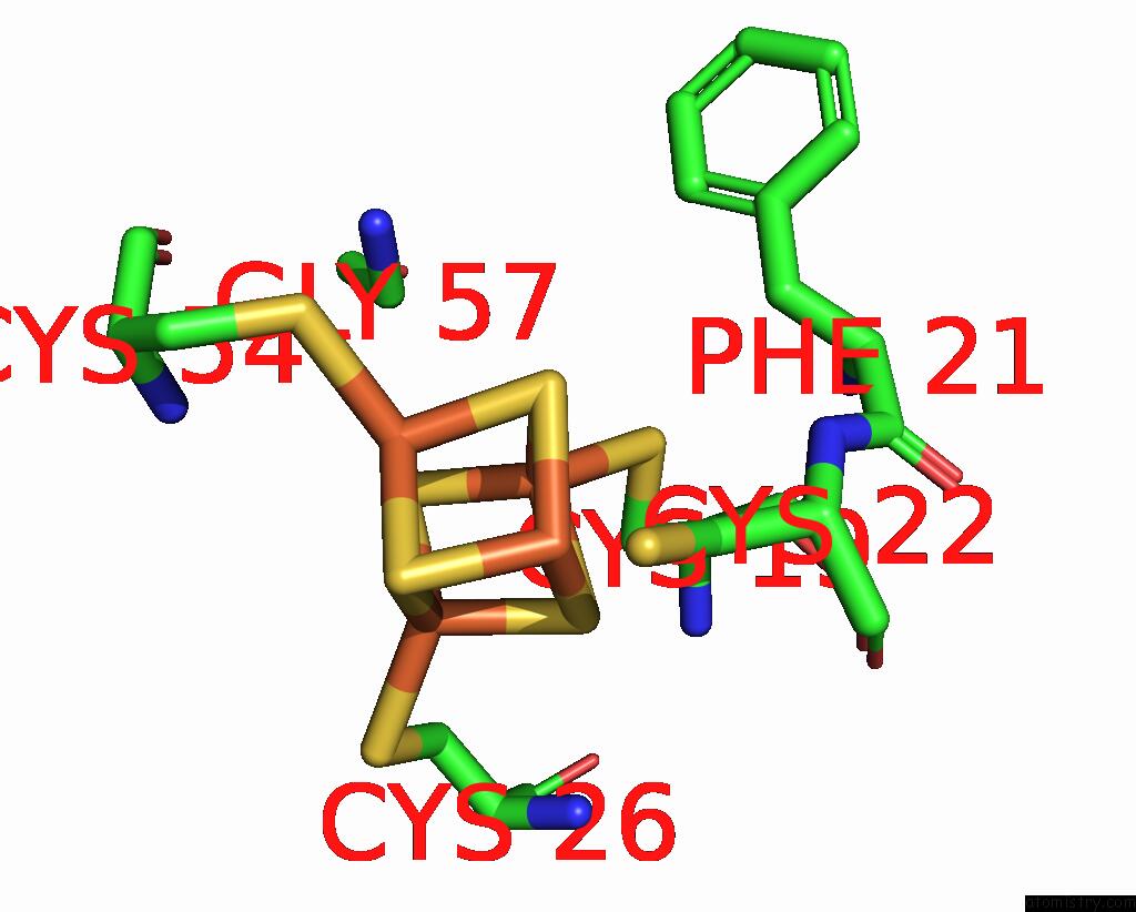
Iron binding site 1 out of 48 in 1ogy

Go back to Iron Binding Sites List in 1ogy
Iron binding site 1 out of 48 in the Crystal Structure of the Heterodimeric Nitrate Reductase From Rhodobacter Sphaeroides


Mono view


Stereo pair view

A full contact list of Iron with other atoms in the Fe binding site number 1 of Crystal Structure of the Heterodimeric Nitrate Reductase From Rhodobacter Sphaeroides within 5.0Å range:
probe atom residue distance (Å) B Occ
A:Fe1801

b:10.0
occ:1.00
FE1 A:SF41801 0.0 10.0 1.0
CB A:CYS22 2.1 10.0 1.0
S4 A:SF41801 2.2 10.5 1.0
S3 A:SF41801 2.2 10.2 1.0
S2 A:SF41801 2.3 12.0 1.0
SG A:CYS22 2.3 12.5 1.0
FE4 A:SF41801 2.6 10.0 1.0
FE2 A:SF41801 2.7 10.0 1.0
FE3 A:SF41801 2.7 10.5 1.0
CA A:CYS22 3.6 10.0 1.0
S1 A:SF41801 3.8 10.0 1.0
N A:CYS22 4.0 11.0 1.0
CB A:THR24 4.1 10.0 1.0
OG1 A:THR24 4.2 10.0 1.0
C A:CYS22 4.4 11.2 1.0
SG A:CYS19 4.6 10.0 1.0
SG A:CYS26 4.7 11.1 1.0
SG A:CYS54 4.7 11.4 1.0
CD A:PRO194 4.8 13.6 1.0
O A:CYS22 4.8 13.2 1.0
N A:THR24 4.9 10.0 1.0

Iron binding site 2 out of 48 in 1ogy

Go back to Iron Binding Sites List in 1ogy
Iron binding site 2 out of 48 in the Crystal Structure of the Heterodimeric Nitrate Reductase From Rhodobacter Sphaeroides


Mono view


Stereo pair view

A full contact list of Iron with other atoms in the Fe binding site number 2 of Crystal Structure of the Heterodimeric Nitrate Reductase From Rhodobacter Sphaeroides within 5.0Å range:
probe atom residue distance (Å) B Occ
A:Fe1801

b:10.0
occ:1.00
FE2 A:SF41801 0.0 10.0 1.0
S3 A:SF41801 2.2 10.2 1.0
S4 A:SF41801 2.2 10.5 1.0
SG A:CYS54 2.3 11.4 1.0
S1 A:SF41801 2.3 10.0 1.0
FE4 A:SF41801 2.7 10.0 1.0
FE3 A:SF41801 2.7 10.5 1.0
FE1 A:SF41801 2.7 10.0 1.0
CB A:CYS54 3.3 10.0 1.0
N A:CYS54 3.8 10.9 1.0
S2 A:SF41801 3.8 12.0 1.0
CA A:CYS54 4.1 10.3 1.0
N A:GLY57 4.5 10.9 1.0
CD A:PRO194 4.6 13.6 1.0
CB A:CYS22 4.7 10.0 1.0
SG A:CYS26 4.7 11.1 1.0
C A:ASN53 4.7 10.4 1.0
SG A:CYS22 4.7 12.5 1.0
SG A:CYS19 4.7 10.0 1.0
CG1 A:ILE195 4.8 11.5 1.0
CA A:GLY57 4.8 10.7 1.0
C A:CYS54 4.8 10.3 1.0
CG A:PRO194 4.8 14.3 1.0
NZ A:LYS56 4.9 11.0 1.0
CB A:PRO194 5.0 14.0 1.0
CA A:ASN53 5.0 10.7 1.0

Iron binding site 3 out of 48 in 1ogy

Go back to Iron Binding Sites List in 1ogy
Iron binding site 3 out of 48 in the Crystal Structure of the Heterodimeric Nitrate Reductase From Rhodobacter Sphaeroides


Mono view


Stereo pair view

A full contact list of Iron with other atoms in the Fe binding site number 3 of Crystal Structure of the Heterodimeric Nitrate Reductase From Rhodobacter Sphaeroides within 5.0Å range:
probe atom residue distance (Å) B Occ
A:Fe1801

b:10.5
occ:1.00
FE3 A:SF41801 0.0 10.5 1.0
SG A:CYS26 2.2 11.1 1.0
S1 A:SF41801 2.2 10.0 1.0
S2 A:SF41801 2.3 12.0 1.0
S4 A:SF41801 2.3 10.5 1.0
FE2 A:SF41801 2.7 10.0 1.0
FE4 A:SF41801 2.7 10.0 1.0
FE1 A:SF41801 2.7 10.0 1.0
CB A:CYS26 3.0 10.0 1.0
S3 A:SF41801 3.8 10.2 1.0
CG1 A:ILE195 4.0 11.5 1.0
N A:CYS26 4.1 10.2 1.0
CA A:CYS26 4.2 10.0 1.0
CB A:ILE195 4.4 12.4 1.0
CB A:THR24 4.6 10.0 1.0
CB A:CYS22 4.6 10.0 1.0
SG A:CYS19 4.6 10.0 1.0
CG2 A:THR24 4.6 10.6 1.0
CB A:CYS19 4.7 10.0 1.0
SG A:CYS22 4.7 12.5 1.0
CA A:ASN53 4.8 10.7 1.0
SG A:CYS54 4.9 11.4 1.0
N A:CYS54 4.9 10.9 1.0

Iron binding site 4 out of 48 in 1ogy

Go back to Iron Binding Sites List in 1ogy
Iron binding site 4 out of 48 in the Crystal Structure of the Heterodimeric Nitrate Reductase From Rhodobacter Sphaeroides


Mono view


Stereo pair view

A full contact list of Iron with other atoms in the Fe binding site number 4 of Crystal Structure of the Heterodimeric Nitrate Reductase From Rhodobacter Sphaeroides within 5.0Å range:
probe atom residue distance (Å) B Occ
A:Fe1801

b:10.0
occ:1.00
FE4 A:SF41801 0.0 10.0 1.0
S2 A:SF41801 2.1 12.0 1.0
S1 A:SF41801 2.1 10.0 1.0
S3 A:SF41801 2.1 10.2 1.0
SG A:CYS19 2.2 10.0 1.0
FE1 A:SF41801 2.6 10.0 1.0
FE2 A:SF41801 2.7 10.0 1.0
FE3 A:SF41801 2.7 10.5 1.0
CB A:CYS19 3.1 10.0 1.0
S4 A:SF41801 3.7 10.5 1.0
CB A:CYS22 3.8 10.0 1.0
CB A:PHE21 4.4 10.0 1.0
CA A:GLY57 4.4 10.7 1.0
SG A:CYS54 4.5 11.4 1.0
CB A:CYS26 4.5 10.0 1.0
N A:CYS22 4.6 11.0 1.0
SG A:CYS26 4.6 11.1 1.0
CA A:CYS19 4.6 10.0 1.0
N A:GLY57 4.7 10.9 1.0
SG A:CYS22 4.8 12.5 1.0
CD1 A:PHE21 4.9 10.8 1.0
CA A:CYS22 4.9 10.0 1.0
N A:PHE21 5.0 10.0 1.0

Iron binding site 5 out of 48 in 1ogy

Go back to Iron Binding Sites List in 1ogy
Iron binding site 5 out of 48 in the Crystal Structure of the Heterodimeric Nitrate Reductase From Rhodobacter Sphaeroides


Mono view


Stereo pair view

A full contact list of Iron with other atoms in the Fe binding site number 5 of Crystal Structure of the Heterodimeric Nitrate Reductase From Rhodobacter Sphaeroides within 5.0Å range:
probe atom residue distance (Å) B Occ
B:Fe1128

b:15.9
occ:1.00
FE B:HEC1128 0.0 15.9 1.0
ND B:HEC1128 1.9 19.4 1.0
NE2 B:HIS44 1.9 20.9 1.0
NE2 B:HIS62 2.0 11.5 1.0
NB B:HEC1128 2.0 15.2 1.0
NC B:HEC1128 2.0 16.6 1.0
NA B:HEC1128 2.0 15.7 1.0
CE1 B:HIS44 2.8 24.4 1.0
CE1 B:HIS62 2.9 10.7 1.0
C4D B:HEC1128 3.0 22.1 1.0
C1D B:HEC1128 3.0 21.6 1.0
C4A B:HEC1128 3.0 14.7 1.0
C4B B:HEC1128 3.0 14.7 1.0
C4C B:HEC1128 3.0 17.2 1.0
C1C B:HEC1128 3.0 16.3 1.0
C1A B:HEC1128 3.0 16.4 1.0
C1B B:HEC1128 3.0 13.5 1.0
CD2 B:HIS44 3.1 26.5 1.0
CD2 B:HIS62 3.1 10.0 1.0
CHB B:HEC1128 3.4 14.1 1.0
CHD B:HEC1128 3.4 18.9 1.0
CHA B:HEC1128 3.4 19.6 1.0
CHC B:HEC1128 3.4 16.4 1.0
ND1 B:HIS44 4.0 27.3 1.0
ND1 B:HIS62 4.1 11.8 1.0
CG B:HIS44 4.1 28.6 1.0
CG B:HIS62 4.2 11.2 1.0
C2B B:HEC1128 4.2 12.2 1.0
C2D B:HEC1128 4.2 25.2 1.0
C3B B:HEC1128 4.2 12.7 1.0
C2C B:HEC1128 4.3 14.6 1.0
C3C B:HEC1128 4.3 16.6 1.0
C3D B:HEC1128 4.3 26.1 1.0
C3A B:HEC1128 4.3 15.5 1.0
C2A B:HEC1128 4.3 15.9 1.0

Iron binding site 6 out of 48 in 1ogy

Go back to Iron Binding Sites List in 1ogy
Iron binding site 6 out of 48 in the Crystal Structure of the Heterodimeric Nitrate Reductase From Rhodobacter Sphaeroides


Mono view


Stereo pair view

A full contact list of Iron with other atoms in the Fe binding site number 6 of Crystal Structure of the Heterodimeric Nitrate Reductase From Rhodobacter Sphaeroides within 5.0Å range:
probe atom residue distance (Å) B Occ
B:Fe1129

b:15.4
occ:1.00
FE B:HEC1129 0.0 15.4 1.0
NE2 B:HIS102 2.0 10.5 1.0
NE2 B:HIS79 2.0 10.2 1.0
NA B:HEC1129 2.0 15.4 1.0
NB B:HEC1129 2.0 14.0 1.0
ND B:HEC1129 2.0 16.7 1.0
NC B:HEC1129 2.0 16.8 1.0
CE1 B:HIS79 2.8 10.0 1.0
CE1 B:HIS102 2.8 10.6 1.0
C1A B:HEC1129 3.0 17.3 1.0
C4A B:HEC1129 3.0 14.2 1.0
C4B B:HEC1129 3.0 14.0 1.0
C1D B:HEC1129 3.0 18.3 1.0
C4D B:HEC1129 3.0 19.2 1.0
C1B B:HEC1129 3.1 13.6 1.0
C1C B:HEC1129 3.1 17.2 1.0
C4C B:HEC1129 3.1 17.3 1.0
CD2 B:HIS102 3.1 10.0 1.0
CD2 B:HIS79 3.2 11.1 1.0
CHB B:HEC1129 3.4 13.2 1.0
CHA B:HEC1129 3.4 18.8 1.0
CHD B:HEC1129 3.4 18.3 1.0
CHC B:HEC1129 3.4 16.1 1.0
ND1 B:HIS102 4.0 11.6 1.0
ND1 B:HIS79 4.0 10.1 1.0
CG B:HIS102 4.2 10.4 1.0
CG B:HIS79 4.2 11.7 1.0
C2A B:HEC1129 4.2 16.8 1.0
C2B B:HEC1129 4.3 13.8 1.0
C3B B:HEC1129 4.3 13.3 1.0
C3A B:HEC1129 4.3 14.6 1.0
C2D B:HEC1129 4.3 20.2 1.0
C2C B:HEC1129 4.3 17.0 1.0
C3C B:HEC1129 4.3 16.9 1.0
C3D B:HEC1129 4.3 22.5 1.0
NH1 B:ARG95 4.8 12.6 1.0

Iron binding site 7 out of 48 in 1ogy

Go back to Iron Binding Sites List in 1ogy
Iron binding site 7 out of 48 in the Crystal Structure of the Heterodimeric Nitrate Reductase From Rhodobacter Sphaeroides


Mono view


Stereo pair view

A full contact list of Iron with other atoms in the Fe binding site number 7 of Crystal Structure of the Heterodimeric Nitrate Reductase From Rhodobacter Sphaeroides within 5.0Å range:
probe atom residue distance (Å) B Occ
C:Fe1801

b:10.0
occ:1.00
FE1 C:SF41801 0.0 10.0 1.0
S4 C:SF41801 2.1 11.4 1.0
S2 C:SF41801 2.2 12.3 1.0
CB C:CYS22 2.2 24.8 1.0
S3 C:SF41801 2.3 10.0 1.0
SG C:CYS22 2.5 27.4 1.0
FE4 C:SF41801 2.6 10.0 1.0
FE3 C:SF41801 2.7 10.0 1.0
FE2 C:SF41801 2.7 10.3 1.0
CA C:CYS22 3.7 23.7 1.0
S1 C:SF41801 3.8 11.8 1.0
CB C:THR24 4.1 21.3 1.0
N C:CYS22 4.1 23.1 1.0
OG1 C:THR24 4.2 21.7 1.0
C C:CYS22 4.4 24.6 1.0
SG C:CYS19 4.5 18.3 1.0
SG C:CYS26 4.6 19.9 1.0
O C:CYS22 4.7 25.1 1.0
SG C:CYS54 4.7 22.3 1.0
CD C:PRO194 4.7 23.3 1.0
N C:THR24 4.8 23.0 1.0
CG2 C:THR24 4.9 21.3 1.0
N C:GLY25 4.9 22.0 1.0

Iron binding site 8 out of 48 in 1ogy

Go back to Iron Binding Sites List in 1ogy
Iron binding site 8 out of 48 in the Crystal Structure of the Heterodimeric Nitrate Reductase From Rhodobacter Sphaeroides


Mono view


Stereo pair view

A full contact list of Iron with other atoms in the Fe binding site number 8 of Crystal Structure of the Heterodimeric Nitrate Reductase From Rhodobacter Sphaeroides within 5.0Å range:
probe atom residue distance (Å) B Occ
C:Fe1801

b:10.3
occ:1.00
FE2 C:SF41801 0.0 10.3 1.0
S3 C:SF41801 2.2 10.0 1.0
S4 C:SF41801 2.3 11.4 1.0
SG C:CYS54 2.3 22.3 1.0
S1 C:SF41801 2.3 11.8 1.0
FE4 C:SF41801 2.7 10.0 1.0
FE3 C:SF41801 2.7 10.0 1.0
FE1 C:SF41801 2.7 10.0 1.0
CB C:CYS54 3.5 21.8 1.0
N C:CYS54 3.7 22.7 1.0
S2 C:SF41801 3.8 12.3 1.0
CA C:CYS54 4.2 21.2 1.0
N C:GLY57 4.5 14.5 1.0
CD C:PRO194 4.5 23.3 1.0
C C:ASN53 4.6 23.8 1.0
SG C:CYS22 4.7 27.4 1.0
CB C:CYS22 4.7 24.8 1.0
SG C:CYS26 4.8 19.9 1.0
CA C:GLY57 4.8 14.9 1.0
CG C:PRO194 4.8 23.1 1.0
CG1 C:ILE195 4.8 19.6 1.0
C C:CYS54 4.9 19.7 1.0
SG C:CYS19 4.9 18.3 1.0
NZ C:LYS56 4.9 10.0 1.0
CA C:ASN53 5.0 24.2 1.0

Iron binding site 9 out of 48 in 1ogy

Go back to Iron Binding Sites List in 1ogy
Iron binding site 9 out of 48 in the Crystal Structure of the Heterodimeric Nitrate Reductase From Rhodobacter Sphaeroides


Mono view


Stereo pair view

A full contact list of Iron with other atoms in the Fe binding site number 9 of Crystal Structure of the Heterodimeric Nitrate Reductase From Rhodobacter Sphaeroides within 5.0Å range:
probe atom residue distance (Å) B Occ
C:Fe1801

b:10.0
occ:1.00
FE3 C:SF41801 0.0 10.0 1.0
SG C:CYS26 2.2 19.9 1.0
S2 C:SF41801 2.2 12.3 1.0
S1 C:SF41801 2.3 11.8 1.0
S4 C:SF41801 2.3 11.4 1.0
FE4 C:SF41801 2.7 10.0 1.0
FE1 C:SF41801 2.7 10.0 1.0
FE2 C:SF41801 2.7 10.3 1.0
CB C:CYS26 3.0 19.5 1.0
S3 C:SF41801 3.8 10.0 1.0
CG1 C:ILE195 4.0 19.6 1.0
N C:CYS26 4.0 19.2 1.0
CA C:CYS26 4.1 19.3 1.0
CB C:ILE195 4.4 18.4 1.0
CB C:THR24 4.6 21.3 1.0
CG2 C:THR24 4.6 21.3 1.0
CB C:CYS19 4.7 15.1 1.0
SG C:CYS19 4.7 18.3 1.0
CA C:ASN53 4.8 24.2 1.0
CB C:CYS22 4.8 24.8 1.0
SG C:CYS54 4.9 22.3 1.0
N C:CYS54 4.9 22.7 1.0

Iron binding site 10 out of 48 in 1ogy

Go back to Iron Binding Sites List in 1ogy
Iron binding site 10 out of 48 in the Crystal Structure of the Heterodimeric Nitrate Reductase From Rhodobacter Sphaeroides


Mono view


Stereo pair view

A full contact list of Iron with other atoms in the Fe binding site number 10 of Crystal Structure of the Heterodimeric Nitrate Reductase From Rhodobacter Sphaeroides within 5.0Å range:
probe atom residue distance (Å) B Occ
C:Fe1801

b:10.0
occ:1.00
FE4 C:SF41801 0.0 10.0 1.0
S2 C:SF41801 2.1 12.3 1.0
S3 C:SF41801 2.1 10.0 1.0
S1 C:SF41801 2.2 11.8 1.0
SG C:CYS19 2.3 18.3 1.0
FE1 C:SF41801 2.6 10.0 1.0
FE2 C:SF41801 2.7 10.3 1.0
FE3 C:SF41801 2.7 10.0 1.0
CB C:CYS19 3.1 15.1 1.0
S4 C:SF41801 3.7 11.4 1.0
CB C:CYS22 4.0 24.8 1.0
CB C:PHE21 4.4 18.2 1.0
CA C:GLY57 4.5 14.9 1.0
CB C:CYS26 4.5 19.5 1.0
CA C:CYS19 4.5 14.9 1.0
N C:CYS22 4.6 23.1 1.0
SG C:CYS26 4.6 19.9 1.0
SG C:CYS54 4.7 22.3 1.0
N C:GLY57 4.8 14.5 1.0
CD1 C:PHE21 4.9 15.7 1.0
SG C:CYS22 4.9 27.4 1.0
N C:PHE21 4.9 16.9 1.0
CA C:CYS22 5.0 23.7 1.0

Reference:

P.Arnoux, M.Sabaty, J.Alric, B.Frangioni, B.Guigliarelli, J.-M.Adriano, D.Pignol. Structural and Redox Plasticity in the Heterodimeric Periplasmic Nitrate Reductase Nat.Struct.Biol. V. 10 928 2003.
ISSN: ISSN 1072-8368
PubMed: 14528294
DOI: 10.1038/NSB994
Page generated: Sun Dec 13 14:27:11 2020

Last articles

Zn in 8WB0
Zn in 8WAX
Zn in 8WAU
Zn in 8WAZ
Zn in 8WAY
Zn in 8WAV
Zn in 8WAW
Zn in 8WAT
Zn in 8W7M
Zn in 8WD3
© Copyright 2008-2020 by atomistry.com
Home   |    Site Map   |    Copyright   |    Contact us   |    Privacy