Iron »
PDB 1ofj-1ozl »
1oqh »
Iron in PDB 1oqh: Crystal Structure of the R124A Mutant of the N-Lobe Human Transferrin
Protein crystallography data
The structure of Crystal Structure of the R124A Mutant of the N-Lobe Human Transferrin, PDB code: 1oqh
was solved by
H.M.Baker,
Q.-Y.He,
S.K.Brigg,
A.B.Mason,
E.N Baker,
with X-Ray Crystallography technique. A brief refinement statistics is given in the table below:
| Resolution Low / High (Å) | 30.00 / 2.40 |
| Space group | P 21 21 21 |
| Cell size a, b, c (Å), α, β, γ (°) | 43.930, 57.120, 135.250, 90.00, 90.00, 90.00 |
| R / Rfree (%) | 21.9 / 28.8 |
Other elements in 1oqh:
The structure of Crystal Structure of the R124A Mutant of the N-Lobe Human Transferrin also contains other interesting chemical elements:
| Potassium | (K) | 1 atom |
Iron Binding Sites:
The binding sites of Iron atom in the Crystal Structure of the R124A Mutant of the N-Lobe Human Transferrin
(pdb code 1oqh). This binding sites where shown within
5.0 Angstroms radius around Iron atom.
In total only one binding site of Iron was determined in the Crystal Structure of the R124A Mutant of the N-Lobe Human Transferrin, PDB code: 1oqh:
In total only one binding site of Iron was determined in the Crystal Structure of the R124A Mutant of the N-Lobe Human Transferrin, PDB code: 1oqh:
Iron binding site 1 out of 1 in 1oqh
Go back to
Iron binding site 1 out
of 1 in the Crystal Structure of the R124A Mutant of the N-Lobe Human Transferrin
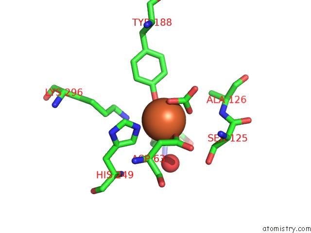
Mono view
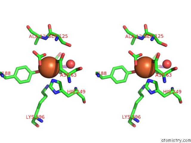
Stereo pair view
Mono view
Stereo pair view
A full contact list of Iron with other atoms in the Fe binding
site number 1 of Crystal Structure of the R124A Mutant of the N-Lobe Human Transferrin within 5.0Å range:
|
Reference:
H.M.Baker,
Q.-Y.He,
S.K.Briggs,
A.B.Mason,
E.N.Baker.
Structural and Functional Consequences of Binding Site Mutations in Transferrin: Crystal Structures of the ASP63GLU and ARG124ALA Mutants of the N-Lobe of Human Transferrin Biochemistry V. 42 7084 2003.
ISSN: ISSN 0006-2960
PubMed: 12795604
DOI: 10.1021/BI020689F
Page generated: Sat Aug 3 12:35:42 2024
ISSN: ISSN 0006-2960
PubMed: 12795604
DOI: 10.1021/BI020689F
Last articles
Zn in 9MJ5Zn in 9HNW
Zn in 9G0L
Zn in 9FNE
Zn in 9DZN
Zn in 9E0I
Zn in 9D32
Zn in 9DAK
Zn in 8ZXC
Zn in 8ZUF