Iron »
PDB 1ofj-1ozl »
1orn »
Iron in PDB 1orn: Structure of A Trapped Endonuclease III-Dna Covalent Intermediate: Estranged-Guanine Complex
Protein crystallography data
The structure of Structure of A Trapped Endonuclease III-Dna Covalent Intermediate: Estranged-Guanine Complex, PDB code: 1orn
was solved by
J.C.Fromme,
G.L.Verdine,
with X-Ray Crystallography technique. A brief refinement statistics is given in the table below:
| Resolution Low / High (Å) | 29.58 / 1.70 |
| Space group | P 21 21 2 |
| Cell size a, b, c (Å), α, β, γ (°) | 67.826, 106.828, 41.708, 90.00, 90.00, 90.00 |
| R / Rfree (%) | 23.5 / 24.8 |
Other elements in 1orn:
The structure of Structure of A Trapped Endonuclease III-Dna Covalent Intermediate: Estranged-Guanine Complex also contains other interesting chemical elements:
| Sodium | (Na) | 1 atom |
Iron Binding Sites:
The binding sites of Iron atom in the Structure of A Trapped Endonuclease III-Dna Covalent Intermediate: Estranged-Guanine Complex
(pdb code 1orn). This binding sites where shown within
5.0 Angstroms radius around Iron atom.
In total 4 binding sites of Iron where determined in the Structure of A Trapped Endonuclease III-Dna Covalent Intermediate: Estranged-Guanine Complex, PDB code: 1orn:
Jump to Iron binding site number: 1; 2; 3; 4;
In total 4 binding sites of Iron where determined in the Structure of A Trapped Endonuclease III-Dna Covalent Intermediate: Estranged-Guanine Complex, PDB code: 1orn:
Jump to Iron binding site number: 1; 2; 3; 4;
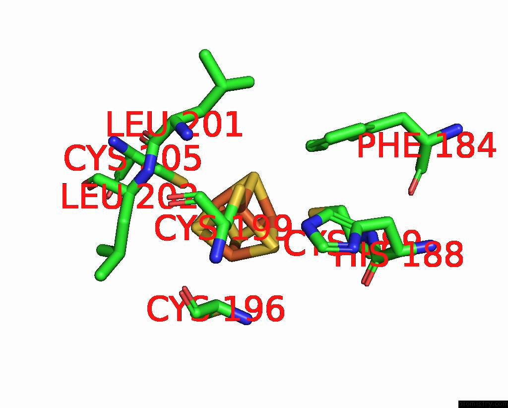
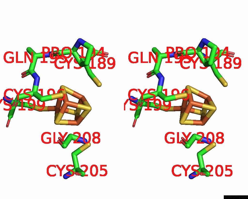
Iron binding site 1 out of 4 in 1orn
Go back to
Iron binding site 1 out
of 4 in the Structure of A Trapped Endonuclease III-Dna Covalent Intermediate: Estranged-Guanine Complex
Mono view

Stereo pair view
Mono view
Stereo pair view
A full contact list of Iron with other atoms in the Fe binding
site number 1 of Structure of A Trapped Endonuclease III-Dna Covalent Intermediate: Estranged-Guanine Complex within 5.0Å range:
|
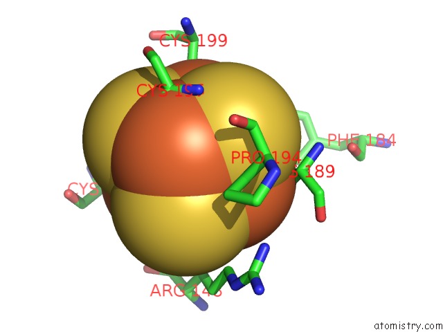
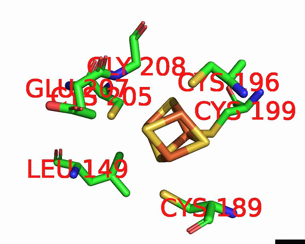
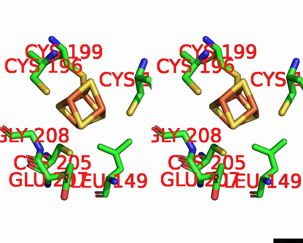
Iron binding site 2 out of 4 in 1orn
Go back to
Iron binding site 2 out
of 4 in the Structure of A Trapped Endonuclease III-Dna Covalent Intermediate: Estranged-Guanine Complex
Mono view
Stereo pair view
Mono view
Stereo pair view
A full contact list of Iron with other atoms in the Fe binding
site number 2 of Structure of A Trapped Endonuclease III-Dna Covalent Intermediate: Estranged-Guanine Complex within 5.0Å range:
|
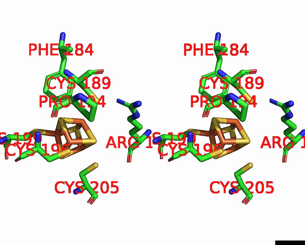
Iron binding site 3 out of 4 in 1orn
Go back to
Iron binding site 3 out
of 4 in the Structure of A Trapped Endonuclease III-Dna Covalent Intermediate: Estranged-Guanine Complex
Mono view
Stereo pair view
Mono view
Stereo pair view
A full contact list of Iron with other atoms in the Fe binding
site number 3 of Structure of A Trapped Endonuclease III-Dna Covalent Intermediate: Estranged-Guanine Complex within 5.0Å range:
|
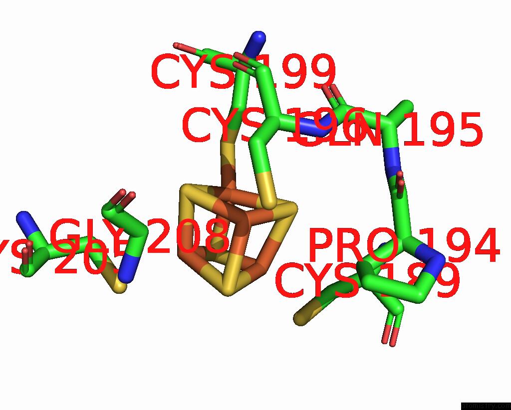
Iron binding site 4 out of 4 in 1orn
Go back to
Iron binding site 4 out
of 4 in the Structure of A Trapped Endonuclease III-Dna Covalent Intermediate: Estranged-Guanine Complex
Mono view
Stereo pair view
Mono view
Stereo pair view
A full contact list of Iron with other atoms in the Fe binding
site number 4 of Structure of A Trapped Endonuclease III-Dna Covalent Intermediate: Estranged-Guanine Complex within 5.0Å range:
|
Reference:
J.C.Fromme,
G.L.Verdine.
Structure of A Trapped Endonuclease III-Dna Covalent Intermediate Embo J. V. 22 3461 2003.
ISSN: ISSN 0261-4189
PubMed: 12840008
DOI: 10.1093/EMBOJ/CDG311
Page generated: Sat Aug 3 12:38:53 2024
ISSN: ISSN 0261-4189
PubMed: 12840008
DOI: 10.1093/EMBOJ/CDG311
Last articles
F in 7R3VF in 7R7K
F in 7QZ9
F in 7QZ8
F in 7R2B
F in 7R26
F in 7QZ6
F in 7QZ2
F in 7QXL
F in 7QZ5