Iron »
PDB 1ofj-1ozl »
1os7 »
Iron in PDB 1os7: Crystal Structure of Taud with Iron, Alpha-Ketoglutarate and Taurine Bound at pH 7.5
Enzymatic activity of Crystal Structure of Taud with Iron, Alpha-Ketoglutarate and Taurine Bound at pH 7.5
All present enzymatic activity of Crystal Structure of Taud with Iron, Alpha-Ketoglutarate and Taurine Bound at pH 7.5:
1.14.11.17;
1.14.11.17;
Protein crystallography data
The structure of Crystal Structure of Taud with Iron, Alpha-Ketoglutarate and Taurine Bound at pH 7.5, PDB code: 1os7
was solved by
J.R.O'brien,
D.J.Schuller,
V.S.Yang,
B.D.Dillard,
W.N.Lanzilotta,
with X-Ray Crystallography technique. A brief refinement statistics is given in the table below:
| Resolution Low / High (Å) | 38.00 / 2.50 |
| Space group | P 21 21 21 |
| Cell size a, b, c (Å), α, β, γ (°) | 92.594, 118.855, 118.832, 90.00, 90.00, 90.00 |
| R / Rfree (%) | 22.4 / 27.8 |
Iron Binding Sites:
The binding sites of Iron atom in the Crystal Structure of Taud with Iron, Alpha-Ketoglutarate and Taurine Bound at pH 7.5
(pdb code 1os7). This binding sites where shown within
5.0 Angstroms radius around Iron atom.
In total 4 binding sites of Iron where determined in the Crystal Structure of Taud with Iron, Alpha-Ketoglutarate and Taurine Bound at pH 7.5, PDB code: 1os7:
Jump to Iron binding site number: 1; 2; 3; 4;
In total 4 binding sites of Iron where determined in the Crystal Structure of Taud with Iron, Alpha-Ketoglutarate and Taurine Bound at pH 7.5, PDB code: 1os7:
Jump to Iron binding site number: 1; 2; 3; 4;
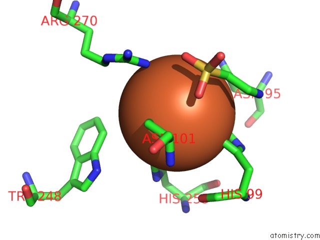
Iron binding site 1 out of 4 in 1os7
Go back to
Iron binding site 1 out
of 4 in the Crystal Structure of Taud with Iron, Alpha-Ketoglutarate and Taurine Bound at pH 7.5
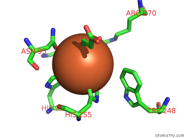
Mono view
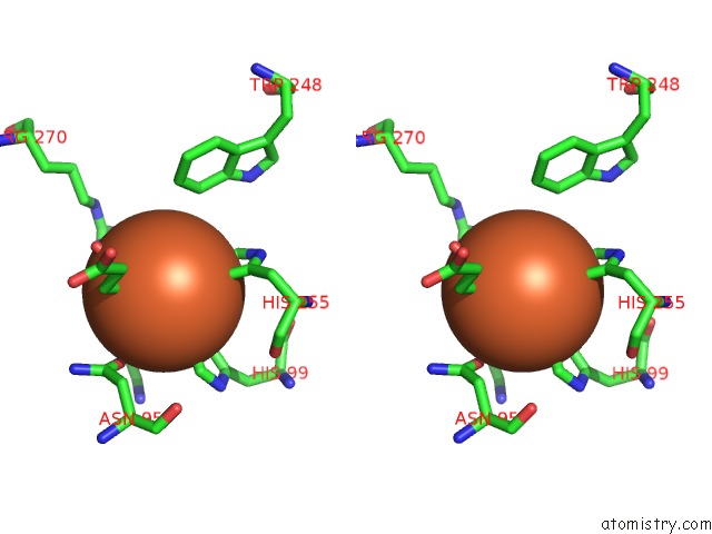
Stereo pair view
Mono view
Stereo pair view
A full contact list of Iron with other atoms in the Fe binding
site number 1 of Crystal Structure of Taud with Iron, Alpha-Ketoglutarate and Taurine Bound at pH 7.5 within 5.0Å range:
|
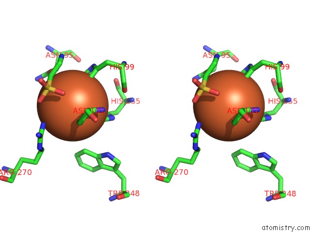
Iron binding site 2 out of 4 in 1os7
Go back to
Iron binding site 2 out
of 4 in the Crystal Structure of Taud with Iron, Alpha-Ketoglutarate and Taurine Bound at pH 7.5
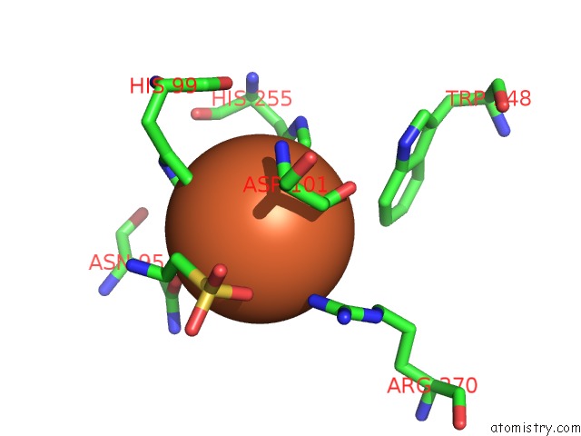
Mono view
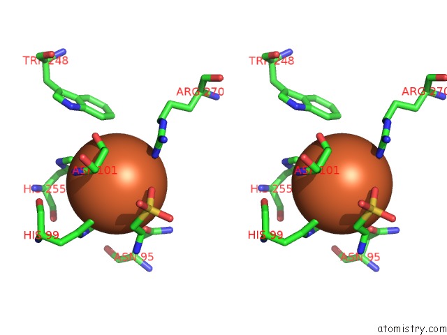
Stereo pair view
Mono view
Stereo pair view
A full contact list of Iron with other atoms in the Fe binding
site number 2 of Crystal Structure of Taud with Iron, Alpha-Ketoglutarate and Taurine Bound at pH 7.5 within 5.0Å range:
|
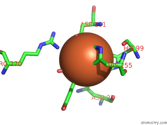
Iron binding site 3 out of 4 in 1os7
Go back to
Iron binding site 3 out
of 4 in the Crystal Structure of Taud with Iron, Alpha-Ketoglutarate and Taurine Bound at pH 7.5
Mono view
Stereo pair view
Mono view
Stereo pair view
A full contact list of Iron with other atoms in the Fe binding
site number 3 of Crystal Structure of Taud with Iron, Alpha-Ketoglutarate and Taurine Bound at pH 7.5 within 5.0Å range:
|
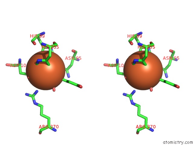
Iron binding site 4 out of 4 in 1os7
Go back to
Iron binding site 4 out
of 4 in the Crystal Structure of Taud with Iron, Alpha-Ketoglutarate and Taurine Bound at pH 7.5
Mono view
Stereo pair view
Mono view
Stereo pair view
A full contact list of Iron with other atoms in the Fe binding
site number 4 of Crystal Structure of Taud with Iron, Alpha-Ketoglutarate and Taurine Bound at pH 7.5 within 5.0Å range:
|
Reference:
J.R.O'brien,
D.J.Schuller,
V.S.Yang,
B.D.Dillard,
W.N.Lanzilotta.
Substrate-Induced Conformational Changes in Escherichia Coli Taurine/Alpha-Ketoglutarate Dioxygenase and Insight Into the Oligomeric Structure Biochemistry V. 42 5547 2003.
ISSN: ISSN 0006-2960
PubMed: 12741810
DOI: 10.1021/BI0341096
Page generated: Sat Aug 3 12:40:54 2024
ISSN: ISSN 0006-2960
PubMed: 12741810
DOI: 10.1021/BI0341096
Last articles
Zn in 9MJ5Zn in 9HNW
Zn in 9G0L
Zn in 9FNE
Zn in 9DZN
Zn in 9E0I
Zn in 9D32
Zn in 9DAK
Zn in 8ZXC
Zn in 8ZUF