Iron »
PDB 1ozr-1pha »
1pah »
Iron in PDB 1pah: Human Phenylalanine Hydroxylase Dimer, Residues 117-424
Enzymatic activity of Human Phenylalanine Hydroxylase Dimer, Residues 117-424
All present enzymatic activity of Human Phenylalanine Hydroxylase Dimer, Residues 117-424:
1.14.16.1;
1.14.16.1;
Protein crystallography data
The structure of Human Phenylalanine Hydroxylase Dimer, Residues 117-424, PDB code: 1pah
was solved by
R.C.Stevens,
H.Erlandsen,
with X-Ray Crystallography technique. A brief refinement statistics is given in the table below:
| Resolution Low / High (Å) | 20.00 / 2.00 |
| Space group | C 2 2 21 |
| Cell size a, b, c (Å), α, β, γ (°) | 66.600, 108.400, 125.700, 90.00, 90.00, 90.00 |
| R / Rfree (%) | 17.6 / 21.8 |
Iron Binding Sites:
The binding sites of Iron atom in the Human Phenylalanine Hydroxylase Dimer, Residues 117-424
(pdb code 1pah). This binding sites where shown within
5.0 Angstroms radius around Iron atom.
In total only one binding site of Iron was determined in the Human Phenylalanine Hydroxylase Dimer, Residues 117-424, PDB code: 1pah:
In total only one binding site of Iron was determined in the Human Phenylalanine Hydroxylase Dimer, Residues 117-424, PDB code: 1pah:
Iron binding site 1 out of 1 in 1pah
Go back to
Iron binding site 1 out
of 1 in the Human Phenylalanine Hydroxylase Dimer, Residues 117-424
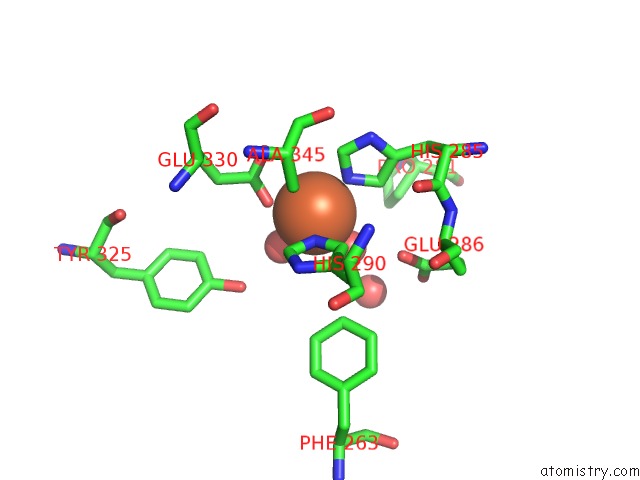
Mono view
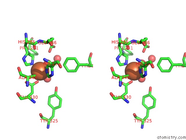
Stereo pair view
Mono view
Stereo pair view
A full contact list of Iron with other atoms in the Fe binding
site number 1 of Human Phenylalanine Hydroxylase Dimer, Residues 117-424 within 5.0Å range:
|
Reference:
H.Erlandsen,
F.Fusetti,
A.Martinez,
E.Hough,
T.Flatmark,
R.C.Stevens.
Crystal Structure of the Catalytic Domain of Human Phenylalanine Hydroxylase Reveals the Structural Basis For Phenylketonuria. Nat.Struct.Biol. V. 4 995 1997.
ISSN: ISSN 1072-8368
PubMed: 9406548
DOI: 10.1038/NSB1297-995
Page generated: Sat Aug 3 13:01:17 2024
ISSN: ISSN 1072-8368
PubMed: 9406548
DOI: 10.1038/NSB1297-995
Last articles
Zn in 9MJ5Zn in 9HNW
Zn in 9G0L
Zn in 9FNE
Zn in 9DZN
Zn in 9E0I
Zn in 9D32
Zn in 9DAK
Zn in 8ZXC
Zn in 8ZUF